
Zhiya Chen, Ying Ji, Wenbo Tang, Mingming Zhang, Yuanyuan Xu, Yawei Wang. Optical Phase Characterization Method for Dynamic Characteristics of Neuronal Discharge[J]. Chinese Journal of Lasers, 2018, 45(6): 0607001
- Chinese Journal of Lasers
- Vol. 45, Issue 6, 0607001 (2018)
![(a) Practical morphology[15] and (b) 3D simplified model of neurons](/richHtml/zgjg/2018/45/6/0607001/img_1.jpg)
Fig. 1. (a) Practical morphology[15] and (b) 3D simplified model of neurons
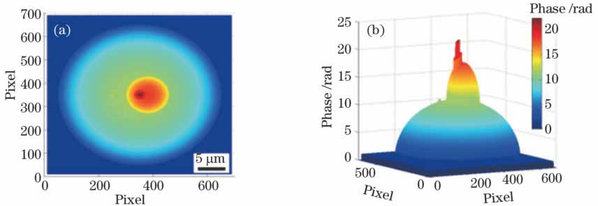
Fig. 2. (a) 2D and (b) 3D unwrapping phases of neuron model along z axis
Fig. 3. Phase of neuron. (a) Gradient distribution; (b) absolute value of gradient; (c) horizontal gradient curve
Fig. 4. Refractive index distribution through live neuron
Fig. 5. (a)-(h) Phases with neuron refractive indexes in range of 1.356-1.367; (i) relationship between phase value of monitoring point and refractive index
Fig. 6. (a) Model, (b) wrapped phase, and (c) unwrapped phase of neuron before compensation; (d) model, (e) wrapped phase, and (f) unwrapped phase of neuron after compensation
Fig. 7. Nucleus and mitochondria in sample. (a) Unwrapped phase; (b) optical thickness; (c) physical thickness; (d) verification of predicted result
Fig. 8. Nucleus and mitochondria of neuronal substructure. (a)-(h) Phases with refractive index variation of cytoplasm; (i) relationship between phase value of monitoring point and difference of refractive index
|
Table 1. Distance between phase gradient mutation points of neuron model in incident direction

Set citation alerts for the article
Please enter your email address
© Copyright 2018-2021 | Chinese Laser Press. All Rights Reserved 沪ICP备15018463号-20