• Chinese Journal of Lasers
  • Vol. 47, Issue 12, 1207006 (2020)
Wang Cheng1, Liu Bin1, Zhou Chu2, Li Nianning1, Zhang Haonan2, Xiang Huazhong1, Zheng Gang1, Wang Xiuli2, and Zhang Dawei3
Author Affiliations
  • 1Key Laboratory of Medical Optical Technology and Instruments, Ministry of Education, Institute of Biomedical Optics and Optometry, University of Shanghai for Science and Technology, Shanghai 200093, China
  • 2Institute of Photomedicine, Shanghai Skin Disease Hospital, Tongji University School of Medicine, Shanghai 200443, China
  • 3Engineering Research Center of Optical Instrument and System, Ministry of Education, Key Laboratory of Modern Optical Systems, University of Shanghai for Science and Technology, Shanghai 200093, China
  • show less
    DOI: 10.3788/CJL202047.1207006 Cite this Article Set citation alerts
    Wang Cheng, Liu Bin, Zhou Chu, Li Nianning, Zhang Haonan, Xiang Huazhong, Zheng Gang, Wang Xiuli, Zhang Dawei. Multispectral Microimaging System with Narrowband LED Illumination[J]. Chinese Journal of Lasers, 2020, 47(12): 1207006 Copy Citation Text show less
    Optical schematic diagram of the system
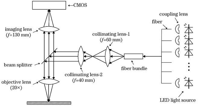
    Fig. 1. Optical schematic diagram of the system
    Picture of the experimental prototype
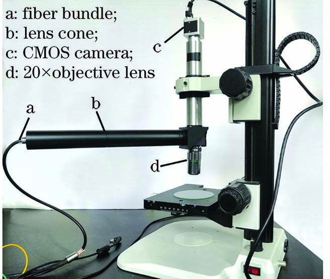
    Fig. 2. Picture of the experimental prototype
    LED emission spectrum
    Fig. 3. LED emission spectrum
    Coupling design
    Fig. 4. Coupling design
    Microscope graticules
    Fig. 5. Microscope graticules
    USAF1951 resolution test target (group 7--6, 228lp/mm)
    Fig. 6. USAF1951 resolution test target (group 7--6, 228lp/mm)
    Single-band images of cancer tissue. (a) 480nm; (b) 520nm; (c) 600nm; (d) 660nm
    Fig. 7. Single-band images of cancer tissue. (a) 480nm; (b) 520nm; (c) 600nm; (d) 660nm
    Single-band images of normal tissue. (a) 480nm; (b) 520nm; (c) 600nm; (d) 660nm
    Fig. 8. Single-band images of normal tissue. (a) 480nm; (b) 520nm; (c) 600nm; (d) 660nm
    False color image and spectra of cancer tissue
    Fig. 9. False color image and spectra of cancer tissue
    False color image and spectra of normal tissue
    Fig. 10. False color image and spectra of normal tissue
    SurfaceCurvature /mmThickness /mmGlassSemi-diameter /mm
    OBJStandardinfinity160
    1Standard32.333SK1112
    2Standard-1052SF1912
    3Standardinfinity12912
    4Standard3058
    5Standard-803SK118
    6Standard-9097.47SF198
    IMAStandardinfinity5
    Table 1. Coupling design parameters
    Group12345678910Average
    dnn0.310.350.390.380.430.330.350.320.270.530.37
    dcc0.220.380.250.360.40.350.280.420.310.350.33
    dnc1.241.021.310.960.990.941.471.191.151.111.14
    Table 2. Sample spectral difference
    Wang Cheng, Liu Bin, Zhou Chu, Li Nianning, Zhang Haonan, Xiang Huazhong, Zheng Gang, Wang Xiuli, Zhang Dawei. Multispectral Microimaging System with Narrowband LED Illumination[J]. Chinese Journal of Lasers, 2020, 47(12): 1207006
    Download Citation