• Acta Physica Sinica
  • Vol. 68, Issue 23, 237801-1 (2019)
Fan Meng1、*, Jin-Hua Hu2, Hui Wang1, Ge-Yin Zou3, Jian-Gong Cui4, and Yue Zhao5、*
Author Affiliations
  • 1School of Information Science and Technology, Shijiazhuang Tiedao University, Shijiazhuang 050043, China
  • 2School of Information and Electrical Engineering, Hebei University of Engineering, Handan 056038, China
  • 3Training Department, Shijiazhuang Post and Telecommunication Technical College, Shijiazhuang 050021, China
  • 4State Key Laboratory of Dynamics Testing Technology, North University of China, Taiyuan 030051, China
  • 5Department of Gastroenterology and Hepatology, The Fourth Hospital of Hebei Medical University, Shijiazhuang 050011, China
  • show less
    DOI: 10.7498/aps.68.20191121 Cite this Article
    Fan Meng, Jin-Hua Hu, Hui Wang, Ge-Yin Zou, Jian-Gong Cui, Yue Zhao. Fluorescence enhancement of monolayer MoS2 in plasmonic resonator [J]. Acta Physica Sinica, 2019, 68(23): 237801-1 Copy Citation Text show less
    The MoS2-cavity coupled system’s (a) structural diagram, (b) scanning electron microscope image, and (c) optical image.MoS2与谐振腔耦合系统(样品)的(a)结构示意图, (b)扫描电子显微镜(SEM)图像和(c)光学图像
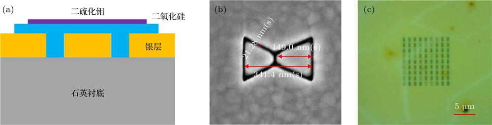
    Fig. 1. The MoS2-cavity coupled system’s (a) structural diagram, (b) scanning electron microscope image, and (c) optical image. MoS2与谐振腔耦合系统(样品)的(a)结构示意图, (b)扫描电子显微镜(SEM)图像和(c)光学图像
    (a) The con-focal microscope setup of measuring PL enhancement of monolayer MoS2; (b) the sample's far-field PL intensity image of EMCCD.(a)研究单层MoS2 PL增强效应的共焦显微系统装置图; (b) EMCCD得到的样品远场PL强度扫描图
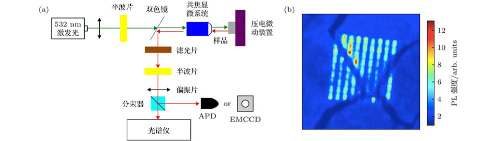
    Fig. 2. (a) The con-focal microscope setup of measuring PL enhancement of monolayer MoS2; (b) the sample's far-field PL intensity image of EMCCD. (a)研究单层MoS2 PL增强效应的共焦显微系统装置图; (b) EMCCD得到的样品远场PL强度扫描图
    The APD scanning images of MoS2 PL enhancement when the angles between the excitation light and resonator’s long-axis are (a) Φ = 0° and (b) Φ = 90°.MoS2辐射光偏振与谐振腔长轴方向在不同夹角 (a) Φ = 0° 和(b) Φ = 90°下得到的PL增强扫描图
    Fig. 3. The APD scanning images of MoS2 PL enhancement when the angles between the excitation light and resonator’s long-axis are (a) Φ = 0° and (b) Φ = 90°. MoS2辐射光偏振与谐振腔长轴方向在不同夹角 (a) Φ = 0° 和(b) Φ = 90°下得到的PL增强扫描图
    The photon counts of APD at different angle combinations of the excitation and detection lights: (a) Φex(co) = 0°; (b) Φex(co) = 90°.不同激发光(探测光)偏振角度下, 探测光(激发光)的光子数变化规律曲线 (a) Φex(co) = 0°; (b) Φex(co) = 90°
    Fig. 4. The photon counts of APD at different angle combinations of the excitation and detection lights: (a) Φex(co) = 0°; (b) Φex(co) = 90°. 不同激发光(探测光)偏振角度下, 探测光(激发光)的光子数变化规律曲线 (a) Φex(co) = 0°; (b) Φex(co) = 90°
    (a) The PL spectra of monolayer MoS2 in different cases; (b) the transmission spectrum of the plasmonic resonator; (c) the PL enhancement of the MoS2-cavity coupled system.(a)单层MoS2在不同情形下的PL谱线; (b)等离子体谐振腔的传输谱; (c)实验中得到的最大PL增强倍数曲线
    Fig. 5. (a) The PL spectra of monolayer MoS2 in different cases; (b) the transmission spectrum of the plasmonic resonator; (c) the PL enhancement of the MoS2-cavity coupled system. (a)单层MoS2在不同情形下的PL谱线; (b)等离子体谐振腔的传输谱; (c)实验中得到的最大PL增强倍数曲线
    Fan Meng, Jin-Hua Hu, Hui Wang, Ge-Yin Zou, Jian-Gong Cui, Yue Zhao. Fluorescence enhancement of monolayer MoS2 in plasmonic resonator [J]. Acta Physica Sinica, 2019, 68(23): 237801-1
    Download Citation