• Chinese Journal of Lasers
  • Vol. 51, Issue 9, 0907015 (2024)
Shuo Liu1、2, Jiang Zhu1、2、*, Xudong Chen1、2, Chongyang Wang1、2, Zongqing Ma1、2, Xiaochen Meng1、2, and Fan Fan1、2
Author Affiliations
  • 1School of Instrument Science and Opto-Electronics Engineering, Beijing Information Science and Technology University, Beijing 102206, China
  • 2Key Laboratory of Ministry of Education for Optoelectronic Measurement Technology and Instruments, Beijing Information Science and Technology University, Beijing 102206, China
  • show less
    DOI: 10.3788/CJL231442 Cite this Article Set citation alerts
    Shuo Liu, Jiang Zhu, Xudong Chen, Chongyang Wang, Zongqing Ma, Xiaochen Meng, Fan Fan. Video‑Guided Handheld High‑Speed Optical Coherence Tomography System[J]. Chinese Journal of Lasers, 2024, 51(9): 0907015 Copy Citation Text show less
    Schematics of the video-guided handheld high-speed optical coherence tomography (OCT) system and the scanning light path in the benchtop OCT system. (a) Schematic of SS-OCT main system ; (b) handheld probe model; (c) schematic of internal light path of handheld probe; (d) schematic of benchtop OCT scanning light path
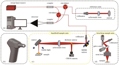
    Fig. 1. Schematics of the video-guided handheld high-speed optical coherence tomography (OCT) system and the scanning light path in the benchtop OCT system. (a) Schematic of SS-OCT main system ; (b) handheld probe model; (c) schematic of internal light path of handheld probe; (d) schematic of benchtop OCT scanning light path
    Imaging of the porcine cornea and tooth by the handheld OCT system. (a) Image of the porcine cornea captured by a cell phone; (b) image of the porcine cornea captured by the video camera in the handheld OCT system; (c) single B-scan image of the porcine cornea captured by the handheld OCT system; (d) image of the porcine tooth captured by a cell phone; (e) image of the porcine tooth captured by the video camera in the handheld OCT system; (f) single B-scan image of the porcine tooth captured by the handheld OCT system
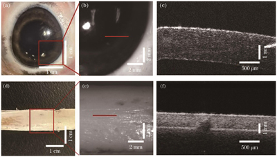
    Fig. 2. Imaging of the porcine cornea and tooth by the handheld OCT system. (a) Image of the porcine cornea captured by a cell phone; (b) image of the porcine cornea captured by the video camera in the handheld OCT system; (c) single B-scan image of the porcine cornea captured by the handheld OCT system; (d) image of the porcine tooth captured by a cell phone; (e) image of the porcine tooth captured by the video camera in the handheld OCT system; (f) single B-scan image of the porcine tooth captured by the handheld OCT system
    Directly averaged and post-registration averaged OCT images of the porcine cornea and tooth (the size of the corneal image is 3 mm×5 mm, and the size of tooth image is 3 mm×7 mm). (a) Directly averaged B-scan image of the porcine cornea; (b) post-registration averaged B-scan image of the porcine cornea; (c) directly averaged B-scan image of the porcine tooth; (d) post-registration averaged B-scan image of the porcine tooth
    Fig. 3. Directly averaged and post-registration averaged OCT images of the porcine cornea and tooth (the size of the corneal image is 3 mm×5 mm, and the size of tooth image is 3 mm×7 mm). (a) Directly averaged B-scan image of the porcine cornea; (b) post-registration averaged B-scan image of the porcine cornea; (c) directly averaged B-scan image of the porcine tooth; (d) post-registration averaged B-scan image of the porcine tooth
    Images of ex-vivo porcine tooth captured by benchtop OCT and handheld OCT systems (the size of the tooth image is 3 mm×7 mm). (a) Single B-scan image captured by the benchtop OCT system; (b) single B-scan image captured by the handheld OCT system; (c) multiple B-scan post-registration averaged OCT image captured by the benchtop OCT system; (d) multiple B-scan post-registration averaged OCT image captured by the handheld OCT system
    Fig. 4. Images of ex-vivo porcine tooth captured by benchtop OCT and handheld OCT systems (the size of the tooth image is 3 mm×7 mm). (a) Single B-scan image captured by the benchtop OCT system; (b) single B-scan image captured by the handheld OCT system; (c) multiple B-scan post-registration averaged OCT image captured by the benchtop OCT system; (d) multiple B-scan post-registration averaged OCT image captured by the handheld OCT system
    Shuo Liu, Jiang Zhu, Xudong Chen, Chongyang Wang, Zongqing Ma, Xiaochen Meng, Fan Fan. Video‑Guided Handheld High‑Speed Optical Coherence Tomography System[J]. Chinese Journal of Lasers, 2024, 51(9): 0907015
    Download Citation