In recent years, lead halide perovskite scintillators have received extensive attention in the field of X-ray imaging. Hard X-ray medical imaging in energy range of 20~120 keV using scintillator detectors, sensitivity and imaging spatial resolution are important performance indicators.
This study aims to explore X-ray imaging property of halide lead perovskite scintillators by simulation.
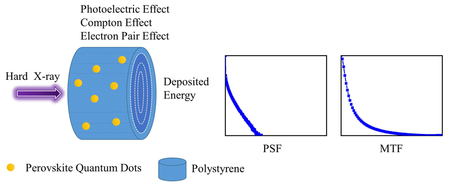
First of all, 3D MAPbBr 3 quantum dots/polystyrene and 2D PEA 2PbBr 4 quantum dots/polystyrene scintillators were taken as research objects. Then, simulation code Geant4 was employed to establish detector model and simulate the X-ray relative detection efficiency and imaging spatial resolution of lead halide perovskite quantum dots/polymer composite scintillators. Finally, the effect of energy and the ratio of perovskite quantum dot occupation on the resolution were explained by secondary electron motion.
The results show that increasing the thickness of the composite scintillator and the proportion of perovskite quantum dots can improve the relative detection efficiency whilst reducing the thickness and increasing the proportion of perovskite quantum dots can improve the spatial resolution. When the absorption efficiency reaches 99.5%, 80% of 3D MAPbBr 3 quantum dots/polystyrene excited by 20 keV X ray obtain the same spatial resolution of 10 lp·mm-1 as CsI. When the incident energy increases to 50 keV, the spatial resolution of CsI is 8 lp·mm-1, while that of lead halide perovskite scintillators is less than 4 lp·mm-1.
It is shown by this study that lead halide perovskites have certain application potential in 20 keV low-energy X-ray medical imaging.