• Chinese Optics Letters
  • Vol. 21, Issue 1, 013401 (2023)
Young-Sung Park1、2, Jieun Hong1, and Jaeho Choi1、*
Author Affiliations
  • 1Photonics Laboratory, Department of Physics, Dankook University, Cheonan 31116, Republic of Korea
  • 2Center for Basic Science Research at DKU, Dankook University, Cheonan 31116, Republic of Korea
  • show less
    DOI: 10.3788/COL202321.013401 Cite this Article Set citation alerts
    Young-Sung Park, Jieun Hong, Jaeho Choi. X-ray volumetric quantitative phase imaging by Foucault differential filtering with linear scanning[J]. Chinese Optics Letters, 2023, 21(1): 013401 Copy Citation Text show less
    Conceptual diagram of the scheme of rendering sectioning images in the experimental setup of FDF. The incident X-ray beams (orange color) from the pinhole array lens (not depicted) pass through the specimen. A and B are objects inside the specimen. k1 and k2, the elements of the Foucault knife-edge (FKA). zo, the location of the objects. Pyz (yi, zj) is the position of the FKA element in terms of the y–z coordinates. The green dot and the green arrow represent the initial position of the scanning and the direction of the FKA movement, respectively.
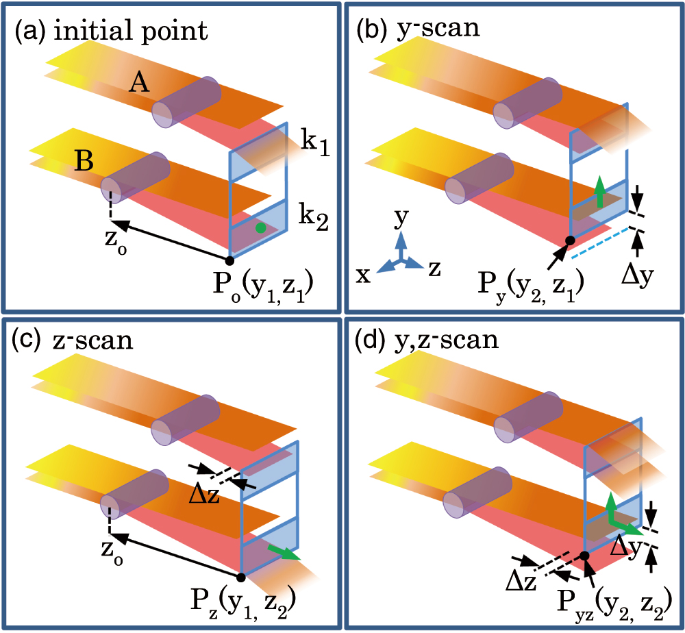
    Fig. 1. Conceptual diagram of the scheme of rendering sectioning images in the experimental setup of FDF. The incident X-ray beams (orange color) from the pinhole array lens (not depicted) pass through the specimen. A and B are objects inside the specimen. k1 and k2, the elements of the Foucault knife-edge (FKA). zo, the location of the objects. Pyz (yi, zj) is the position of the FKA element in terms of the yz coordinates. The green dot and the green arrow represent the initial position of the scanning and the direction of the FKA movement, respectively.
    Images of the gills of a Poecilia reticulata fish. (a) The optical microscopic image. (b) The X-ray absorption image of the gills. (c) Example of the X-ray quantitative phase images in the xy plane. 1, filaments; 2, arches; 3, anchors. The inset scale bar is 200 µm.
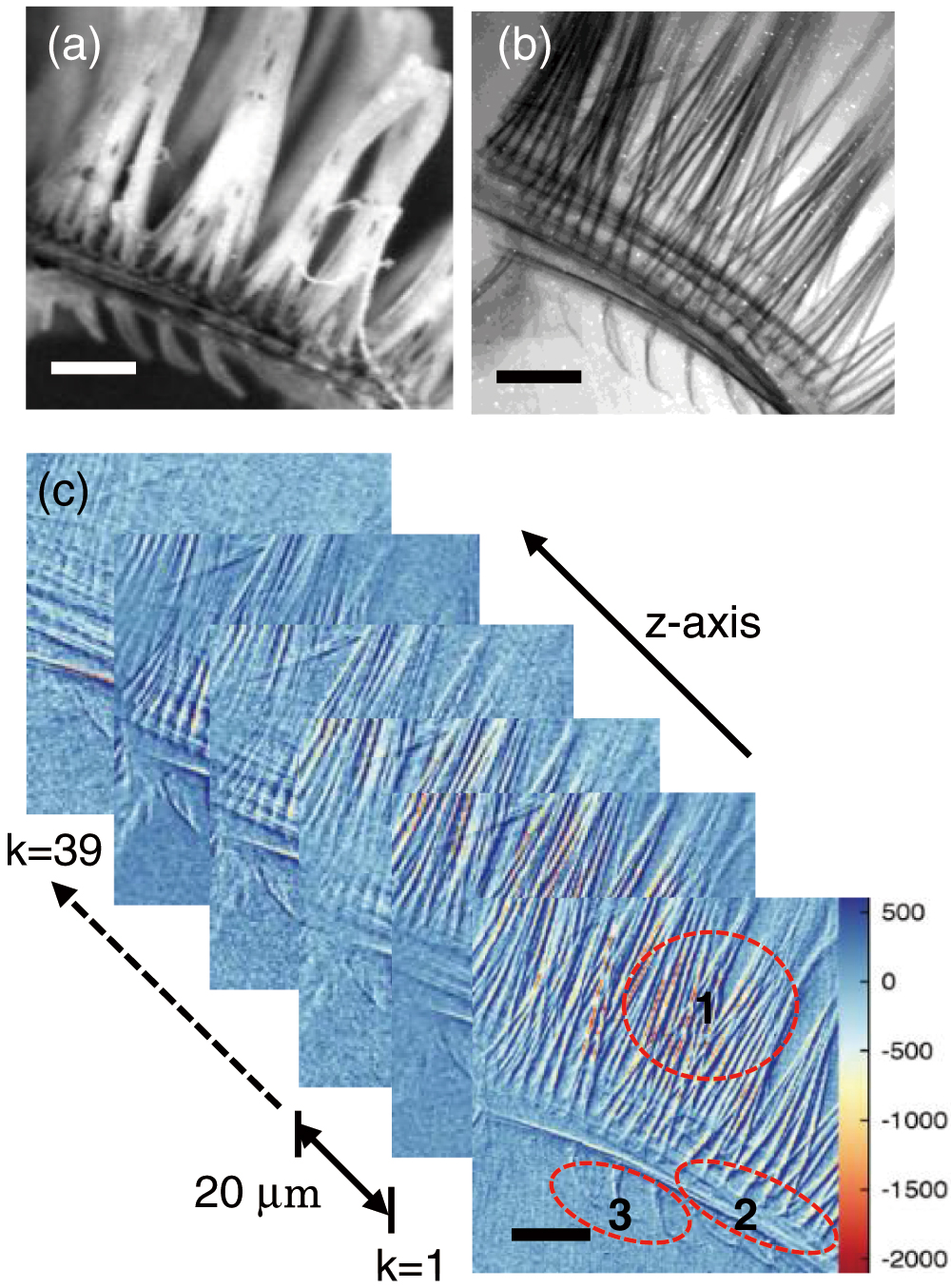
    Fig. 2. Images of the gills of a Poecilia reticulata fish. (a) The optical microscopic image. (b) The X-ray absorption image of the gills. (c) Example of the X-ray quantitative phase images in the xy plane. 1, filaments; 2, arches; 3, anchors. The inset scale bar is 200 µm.
    Volumetric image by the stacked 39-layer xy-plane XQPI. (a) Full-scale view of the 3D rendering image. (b) The cross-sectional planes were perpendicular to the longitudinal direction of filaments. (c) The cut view along the filament direction. The node (the green circle) is shown.
    Fig. 3. Volumetric image by the stacked 39-layer xy-plane XQPI. (a) Full-scale view of the 3D rendering image. (b) The cross-sectional planes were perpendicular to the longitudinal direction of filaments. (c) The cut view along the filament direction. The node (the green circle) is shown.
    Region of interest (ROI) in the volumetric image. (a) The yellow box is the represented ROI on the xy plane of the volumetric XQPI. (b) The white dot lines (L, M) in the enlarged view of ROI are the cut lines for (c) and (d). The red boxes in (c) and (d) show the cross-section views cut through to the z axis along the dot lines L and M. (c) The mucus layers were visualized in between the filaments. The cross-sectional view of the node on the filament end is shown. (d) The connections and nodes between the arch and the filament are shown.
    Fig. 4. Region of interest (ROI) in the volumetric image. (a) The yellow box is the represented ROI on the xy plane of the volumetric XQPI. (b) The white dot lines (L, M) in the enlarged view of ROI are the cut lines for (c) and (d). The red boxes in (c) and (d) show the cross-section views cut through to the z axis along the dot lines L and M. (c) The mucus layers were visualized in between the filaments. The cross-sectional view of the node on the filament end is shown. (d) The connections and nodes between the arch and the filament are shown.
    Young-Sung Park, Jieun Hong, Jaeho Choi. X-ray volumetric quantitative phase imaging by Foucault differential filtering with linear scanning[J]. Chinese Optics Letters, 2023, 21(1): 013401
    Download Citation