
Harry Miyosi Silalahi, Wei-Fan Chiang, Yi-Hong Shih, Wan-Yi Wei, Jou-Yu Su, Chia-Yi Huang, "Folding metamaterials with extremely strong electromagnetic resonance," Photonics Res. 10, 2215 (2022)
- Photonics Research
- Vol. 10, Issue 9, 2215 (2022)
Abstract
1. INTRODUCTION
Terahertz waves have attracted much attention due to their unique physical properties, and have been widely used in many fields, such as communications, imaging, and biomedical applications. Terahertz waves have benefits in biomedical applications [1–5]. With large wavelengths and low photon energies, they can be used to detect live human bodies and biomedical samples via their perspective images, so the detection is a nondestructive and non-ionizing method. Terahertz waves can distinguish organic macromolecules because their specific absorptions from molecular rotation, molecular vibration, hydrogen bonding, and van der Waals interaction are mostly in the terahertz region.
Metamaterials comprising split-ring resonators (SRRs) are promising artificial materials because of their abilities to manipulate electromagnetic waves [6–10] and sense dielectric materials [11–14]. Such abilities arise from their electromagnetic resonance. Metamaterials absorb incident electromagnetic waves at their resonance frequencies, reducing their transmittances at the frequencies. Metamaterials with strong electromagnetic resonance are fascinating due to their high contrast ratios, quality factors, and refractive index sensitivities. Such metamaterials have potential in biomedical sensing.
Water can be used to dilute, preserve, and disperse live biomedical samples. Therefore, reflective terahertz metamaterials have been used to sense water-based biomedical samples [15,16]. However, transmissive terahertz metamaterials cannot detect water-based biomedical samples because water strongly absorbs terahertz waves [17]. Transmissive terahertz metamaterials are easier to integrate terahertz emitters and receivers into single and compact devices than reflective terahertz metamaterials. Therefore, the detection of water-based biomedical samples using transmissive terahertz metamaterials is a big challenge.
Sign up for Photonics Research TOC. Get the latest issue of Photonics Research delivered right to you!Sign up now
This work fabricates a transmissive terahertz metamaterial using a folding metamaterial. The folding metamaterial is made by increasing the height of the SRRs of a metamaterial to 20.7 μm and then removing their bulks, leaving SRRs with nano-profiles. The folding metamaterial has an experimental resonance transmittance and quality factor of
2. MATERIALS AND METHODS
Figure 1(a) presents the design of a folding metamaterial, which is made by folding a thin metamaterial. The folding metamaterial comprises SRRs with nano-profiles. A common metamaterial is used to evaluate the electromagnetic resonance of the folding metamaterial. The common metamaterial comprises SRRs without nano-profiles. The fabrications of the folding and common metamaterials are presented in Appendix A.
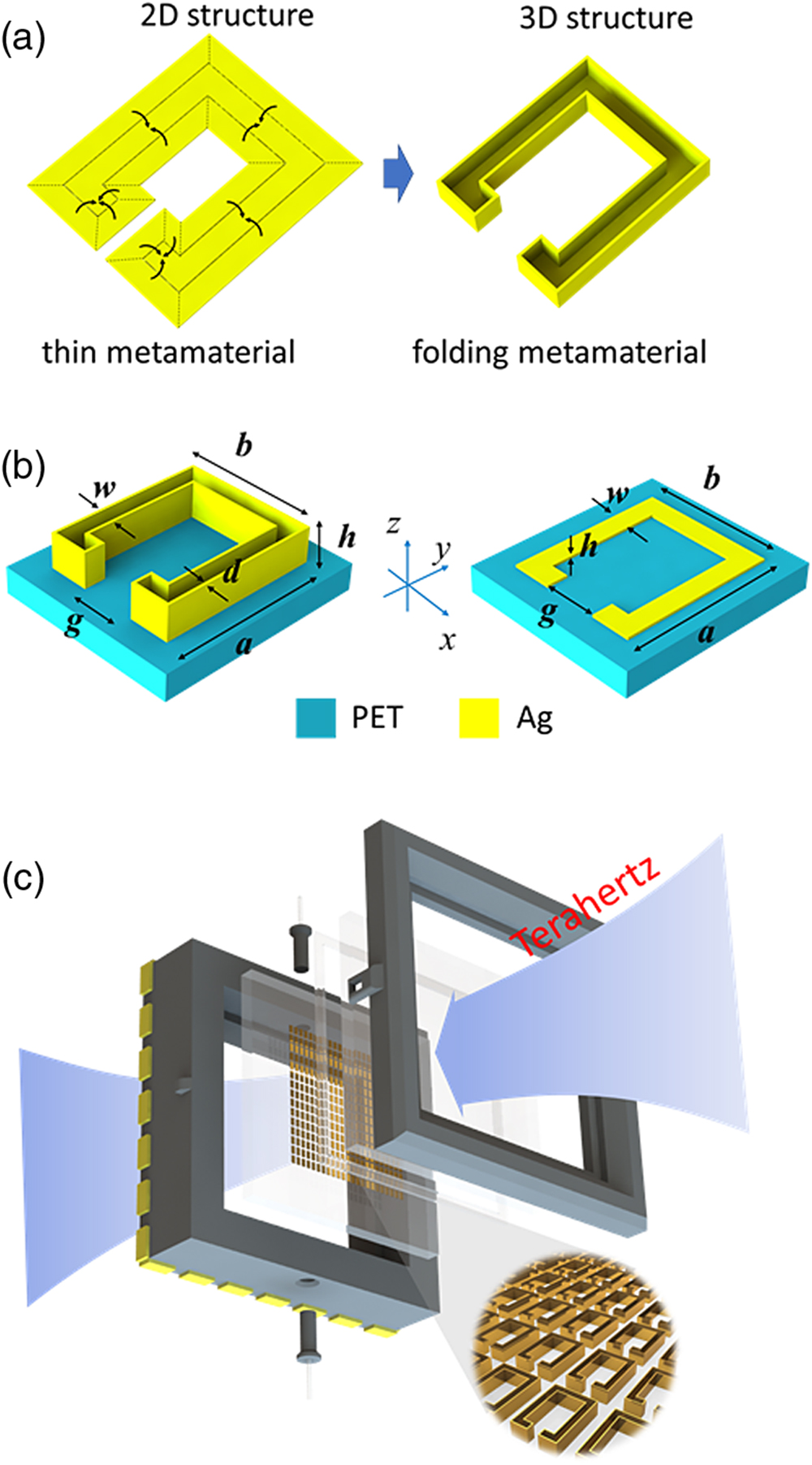
Figure 1.(a) Design of folding metamaterial. The folding metamaterial comprises SRRs with nano-profiles. (b) Geometrical dimensions of folding and common metamaterials. (c) Illustration of biosensor based on folding metamaterial.
Figure 1(b) presents the geometrical dimensions of folding and common metamaterials. The folding metamaterial is designed to have a width (
Figures 2(a) and 2(b) present scanning electron microscope images of the folding metamaterial at 45° and 80° angles of incidence, respectively. The folding metamaterial has a height of 20.7 μm, and the nano-profiles of its SRRs have a width of 500 nm. Therefore, the SRRs in the folding metamaterial have slightly bent nano-profiles due to the high aspect ratio (41.4) of the height (20.7 μm) of the SRRs to the width (500 nm) of their nano-profiles. Figures 2(c) and 2(d) show that the common metamaterial has a height of 500 nm. The folding metamaterial has a larger height than the common metamaterial, so the former has a larger surface area than the latter.
![]()
Figure 2.Scanning electron microscope images of folding metamaterial with height of 20.7 μm at (a) 45° and (b) 80° angles of incidence. Scanning electron microscope images of common metamaterial with height of 500 nm at (c) 45° and (d) 80° angles of incidence.
3. RESULTS AND DISCUSSION
A. Experimental Results
Figure 3 presents the experimental and simulated spectra of the folding and common metamaterials. The spectra are obtained from a terahertz spectrometer (TPS 3000, TeraView) in transmission mode, and the polarized direction of incident terahertz waves is set parallel to the gaps of the SRRs of the folding and common metamaterials. The folding (common) metamaterial has an experimental transmittance of
![]()
Figure 3.(a) Experimental (solid lines) and simulated (dashed lines) spectra of folding and common metamaterials. (b) Surface current distributions of SRRs with and without nano-profiles. (c) Theoretical models of SRRs with and without nano-profiles.
The quality factor of a metamaterial can be used to evaluate the sharpness of its electromagnetic resonance. The quality factor is defined by
The folding (common) metamaterial has an experimental quality factor of 37.0 (11.9) due to its
The transmittance contrast
B. Simulated Results
A simulation is performed using CST software to verify the experimental spectra of the folding and common metamaterials. SRRs with and without nano-profiles are used in the simulation, as presented in Fig. 1(b). The PET substrates of the SRRs have a refractive index of 1.73 in the simulation [11]. Silver has an electrical conductivity of
Figure 3(a) presents the simulated spectra of folding and common metamaterials. Table 1 presents a comparison of the simulated and experimental data of folding and common metamaterials. The folding metamaterial has a larger simulated resonance frequency, smaller simulated resonance transmittance, and larger simulated quality factor than the common metamaterial. Simulated data verify the experimental data in Fig. 3(a). Therefore, the folding metamaterial has stronger electromagnetic resonance than the common metamaterial. The folding metamaterial exhibits a discrepancy between the experimental resonance transmittance and the simulated one, and has a discrepancy between the experimental quality factor and the simulated one. The discrepancies between the experimental data and simulated data arise from the defects [23] and coupling [21,24,25] of the folding metamaterial.
Comparison of Simulated and Experimental Data of Folding and Common Metamaterials
| Type | Resonance Frequency (THz) | Resonance Transmittance (dB) | Quality Factor | |||||
|---|---|---|---|---|---|---|---|---|
| Sim. | Exp. | Sim. | Exp. | Sim. | Exp. | Sim. | Exp. | |
| Folding | 0.790 | 0.776 | −53 | −49 | 0.006 | 0.021 | 131.7 | 37.0 |
| Common | 0.650 | 0.654 | −26 | −25 | 0.008 | 0.055 | 81.3 | 11.9 |
Figure 3(b) presents the surface current distributions of SRRs with and without nano-profiles. As SRRs with and without nano-profiles have electromagnetic resonance, the excited currents flow on their surfaces. An SRR with a nano-profile has a larger surface current than that without a nano-profile because the former has a larger surface area than the latter. Therefore, the folding metamaterial has stronger electromagnetic resonance than common metamaterials.
C. Theoretical Analysis
An SRR can be regarded as a resistor–inductor–capacitor (RLC) circuit in series [26]. The magnitude
Equation (3) reveals that
Substituting
Equation (5) depicts that a small resistance in an SRR decreases its resonance transmittance. In addition, Eq. (2) reveals that the resistance of an SRR can be reduced by increasing its surface area. Therefore, an increase in the surface area of an SRR is crucial to enhance its electromagnetic resonance. This work uses the nano-profile structure with a high aspect ratio of 41.4 to increase the surface areas of SRRs, so the folding metamaterial has extremely strong electromagnetic resonance.
Figure 3(c) presents the theoretical models of SRRs with and without nano-profiles. The SRR with the nano-profile comprises a planar SRR and thin nano-subprofiles. The SRR without a nano-profile is a planar SRR. The two planar SRRs in Fig. 3(c) have the same geometrical dimensions. Suppose the planar SRRs have an identical resistance of
D. Refractive Index Sensitivity
Figure 4(a) displays the near-field distributions of SRRs with and without nano-profiles. An SRR with a nano-profile has a larger hot spot than that without a nano-profile because the former has a larger surface area than the latter. This result reveals that an SRR with a nano-profile has a stronger near field than that without a nano-profile. In other words, an SRR with a nano-profile interacts more with air than that without a nano-profile. Therefore, the folding metamaterial is highly sensitive to dielectric layers that are deposited on them.
![]()
Figure 4.(a) Near-field distributions of SRRs with and without nano-profiles. (b) Experimental spectra of fluidic cells with folding metamaterials at various heights (
The refractive index sensitivity
Figure 4(b) presents the experimental spectra of the fluidic cells with the folding metamaterials at various heights (
A figure of merit (FoM) parameter is interesting to study the sensing performance of the folding metamaterial. The FOM is defined as the ratio of sensitivity (S) and 3 dB-bandwidth (
E. Detection of Rabbit Blood Using Folding Metamaterials
The detection of an aqueous solution using folding and common metamaterials is proposed. The folding and common metamaterials have heights of 20.7 μm and 500 nm, respectively. One of two empty samples is obtained by separating a PET substrate with the folding metamaterial and bare PET substrate using 188-μm-thick plastic spacers, and the other is obtained by separating a PET substrate with the common metamaterial and bare PET substrate using 188-μm-thick plastic spacers. The empty samples are filled with air or rabbit blood. The fluidic samples with the folding and common metamaterials are obtained following the filling.
A rabbit-blood sample without a metamaterial is used to evaluate the performance of the folding metamaterial. An empty sample is made by separating two bare PET substrates using 188-μm-thick plastic spacers, and then is filled with rabbit blood. Blood samples were taken from a healthy rabbit by a qualified clinician. The blood samples were taken and immediately stored in a blood collection tube with an anticoagulant. The blood samples were centrifuged to split them into blood cells and plasma. The plasma was taken out of the blood collection tube using a syringe. Then, the blood cells were injected into folding and common metamaterial cells. The rabbit-blood sample without a metamaterial is obtained following the filling.
Figure 5(a) presents the experimental spectra of the fluidic sample with the folding metamaterial imbedded into the air and rabbit-blood layers. The green curve in Fig. 5(a) is the terahertz spectrum of the rabbit-blood sample without a metamaterial. The rabbit-blood sample without a metamaterial has low transmittances at the frequencies in its terahertz spectrum. This result reveals that the rabbit-blood layer with a thickness of 188 μm in this sample strongly absorbs the incident terahertz waves.
![]()
Figure 5.(a) Experimental spectra of fluidic sample with folding metamaterial imbedded into air and rabbit-blood layers. The green curve is the terahertz spectrum of the rabbit-blood sample without a metamaterial. (b) Experimental spectra of fluidic sample with common metamaterial imbedded into air and rabbit-blood layers.
The black curve in Fig. 5(a) reveals that the fluidic sample with the folding metamaterial imbedded into the air layer has a resonance frequency of 0.788 THz. The resonance frequency arises from the RLC resonance of the folding metamaterial. The transmittance (
Figure 5(b) presents the experimental spectra of the fluidic sample with the common metamaterial imbedded into the air and rabbit-blood layers. The black curve in Fig. 5(b) reveals that the fluidic sample with the common metamaterial imbedded into the air layer has a resonance frequency of 0.655 THz. The transmittance (
The results in Figs. 5(a) and 5(b) reveal that the folding metamaterial detects the rabbit-blood layer with a thickness of 188 μm. Therefore, the folding metamaterial has the potential for detecting the products of live microorganisms with geometrical sizes up to several hundreds of micrometers, such as hydrogen gas, hydrocarbons, and antibodies [39–41].
4. CONCLUSION
The new design, fabrication, characterization, simulation, and application of the folding metamaterial comprising SRRs with nano-profiles that have a high aspect ratio of 41.4 are proposed in this work. The resonance transmittance (
The folding metamaterial exhibits a large refractive index sensitivity (647 GHz/RIU) of the folding metamaterial, and can be used to develop terahertz metamaterials with large refractive index sensitivities. The folding metamaterial has a significant resonance peak as the rabbit-blood layer with a thickness of 188 μm is deposited on it. Therefore, the folding metamaterial has the potential for sensing the products of live microorganisms with geometrical sizes of up to several hundreds of micrometers, such as hydrogen gas, hydrocarbons, and antibodies.
Acknowledgment
Acknowledgment. We thank Dr. Wei-Fan Chiang for his discovery in the fabrication process of folding structures.
APPENDIX A: SAMPLE PREPARATION AND FABRICATION
The folding metamaterial is fabricated using a photolithography, thermal evaporation, and lift-off process as presented in Fig.
![]()
Figure 6.Fabrication of (a) folding and (b) common metamaterials.
A common metamaterial is used to evaluate the electromagnetic resonance of the folding metamaterial. Figure
Folding metamaterials at nanoscale may be fabricated via the proposed method in this work. Such metamaterials are able to manipulate visible light. Folding metamaterials at a nanoscale can be used in biosensing, such as dangerous viruses, body tissues, and cancer cells.
APPENDIX B: TIME-DOMAIN SPECTROSCOPY SYSTEM
All the folding and common metamaterials are measured by using a terahertz spectrometer (TPS 3000, TeraView) in transmission mode, and the polarized direction of incident terahertz waves is set parallel to the gaps of the SRRs of the metamaterials. The infrared light from a femtosecond Ti:sapphire laser (pulse duration:
APPENDIX C: SOLID METAMATERIAL
Figure
![]()
Figure 7.Simulated spectra of the folding and solid metamaterials with a thickness of 20.7 μm. The insets present the simulated models and surface current distributions of folding and solid metamaterials.
A previous work reported that a 9-μm-thick solid metamaterial was fabricated using electroplating [
References
[1] J. Zhang, S. Li, W. Le. Advances of terahertz technology in neuroscience: current status and a future perspective. iScience, 24, 103548(2021).
[2] L. Sun, L. Zhao, R.-Y. Peng. Research progress in the effects of terahertz waves on biomacromolecules. Mil. Med. Res., 8, 28(2021).
[3] Y. Ma, B. Dong, C. Lee. Progress of infrared guided-wave nanophotonic sensors and devices. Nano Converg., 7, 12(2020).
[4] X. Liu, W. Liu, Z. Ren, Y. Ma, B. Dong, G. Zhou, C. Lee. Progress of optomechanical micro/nano sensors: a review. Int. J. Optomechatron., 15, 120-159(2021).
[5] C. Xu, Z. Ren, J. Wei, C. Lee. Reconfigurable terahertz metamaterials: from fundamental principles to advanced 6G applications. iScience, 25, 103799(2022).
[6] W. F. Chiang, H. M. Silalahi, Y. C. Chiang, M. C. Hsu, Y. S. Zhang, J. H. Liu, Y. Yu, C. R. Lee, C. Y. Huang. Continuously tunable intensity modulators with large switching contrasts using liquid crystal elastomer films that are deposited with terahertz metamaterials. Opt. Express, 28, 27676-27687(2020).
[7] P. Pitchappa, C. P. Ho, P. Kropelnicki, N. Singh, D.-L. Kwong, C. Lee. Dual band complementary metamaterial absorber in near infrared region. J. Appl. Phys., 115, 193109(2014).
[8] P. Pitchappa, C. Pei Ho, Y. S. Lin, P. Kropelnicki, C. Y. Huang, N. Singh, C. Lee. Micro-electro-mechanically tunable metamaterial with enhanced electro-optic performance. Appl. Phys. Lett., 104, 151104(2014).
[9] P. Pitchappa, A. Kumar, R. Singh, C. Lee, N. Wang. Terahertz MEMS metadevices. J. Micromech. Microeng., 31, 113001(2021).
[10] A. Kumar, A. Solanki, M. Manjappa, S. Ramesh, Y. K. Srivastava, P. Agarwal, T. C. Sum, R. Singh. Excitons in 2D perovskites for ultrafast terahertz photonic devices. Sci. Adv., 6, eaax8821(2020).
[11] H. M. Silalahi, Y. P. Chen, Y. H. Shih, Y. S. Chen, X. Y. Lin, J. H. Liu, C. Y. Huang. Floating terahertz metamaterials with extremely large refractive index sensitivities. Photon. Res., 9, 1970-1978(2021).
[12] W. F. Chiang, S. X. Lin, Y. X. Lee, Y. H. Shih, J. H. Liu, H. M. Silalahi, C. R. Lee, C. Y. Huang. Effect of thicknesses of liquid crystal layers on shift of resonance frequencies of metamaterials. Coatings, 11, 578(2021).
[13] K. Shih, P. Pitchappa, L. Jin, C.-H. Chen, R. Singh, C. Lee. Nanofluidic terahertz metasensor for sensing in aqueous environment. Appl. Phys. Lett., 113, 071105(2018).
[14] Y.-S. Lin, Z. Xu. Reconfigurable metamaterials for optoelectronic applications. Int. J. Optomechatron., 14, 78-93(2020).
[15] F. Lan, F. Luo, P. Mazumder, Z. Yang, L. Meng, Z. Bao, J. Zhou, Y. Zhang, S. Liang, Z. Shi, A. R. Khan, Z. Zhang, L. Wang, J. Yin, H. Zeng. Dual-band refractometric terahertz biosensing with intense wave-matter-overlap microfluidic channel. Biomed. Opt. Express, 10, 3789-3799(2019).
[16] L. Liang, X. Hu, L. Wen, Y. Zhu, X. Yang, J. Zhou, Y. Zhang, I. E. Carranza, J. Grant, C. Jiang, D. R. S. Cumming, B. Li, Q. Chen. Unity integration of grating slot waveguide and microfluid for terahertz sensing. Laser Photon. Rev., 12, 1800078(2018).
[17] K. Shih, P. Pitchappa, M. Manjappa, C. P. Ho, R. Singh, C. Lee. Microfluidic metamaterial sensor: selective trapping and remote sensing of microparticles. J. Appl. Phys., 121, 023102(2017).
[18] N. R. Han, Z. C. Chen, C. S. Lim, B. Ng, M. H. Hong. Broadband multi-layer terahertz metamaterials fabrication and characterization on flexible substrates. Opt. Express, 19, 6990-6998(2011).
[19] K. Aydin, I. Bulu, K. Guven, M. Kafesaki, C. M. Soukoulis, E. Ozbay. Investigation of magnetic resonances for different split-ring resonator parameters and designs. New J. Phys., 7, 168(2005).
[20] Z. Wang, Z. Geng, W. Fang. Exploring performance of THz metamaterial biosensor based on flexible thin-film. Opt. Express, 28, 26370-26384(2020).
[21] R. Singh, E. Smirnova, A. J. Taylor, J. F. O’Hara, W. Zhang. Optically thin terahertz metamaterials. Opt. Express, 16, 6537-6543(2008).
[22] N. Gneiding, O. Zhuromskyy, E. Shamonina, U. Peschel. Circuit model optimization of a nano split ring resonator dimer antenna operating in infrared spectral range. J. Appl. Phys., 116, 164311(2014).
[23] C. H. Zhang, J. B. Wu, B. B. Jin, Z. M. Ji, L. Kang, W. W. Xu, J. Chen, M. Tonouchi, P. H. Wu. Low-loss terahertz metamaterial from superconducting niobium nitride films. Opt. Express, 20, 42-47(2012).
[24] J. Wu, B. Jin, Y. Xue, C. Zhang, H. Dai, L. Zhang, C. Cao, L. Kang, W. Xu, J. Chen, P. Wu. Tuning of superconducting niobium nitride terahertz metamaterials. Opt. Express, 19, 12021-12026(2011).
[25] R. Singh, A. K. Azad, J. F. O’Hara, A. J. Taylor, W. Zhang. Effect of metal permittivity on resonant properties of terahertz metamaterials. Opt. Lett., 33, 1506-1508(2008).
[26] R. Marqués, F. Mesa, J. Martel, F. Medina. Comparative analysis of edge-and broadside-coupled split ring resonators for metamaterial design-theory and experiments. IEEE Trans. Antennas Propag., 51, 2572-2581(2003).
[27] D. R. Chowdhury, J. F. O’Hara, A. J. Taylor, A. K. Azad. Orthogonally twisted planar concentric split ring resonators towards strong near field coupled terahertz metamaterials. Appl. Phys. Lett., 104, 101105(2014).
[28] R. Cheng, L. Xu, X. Yu, L. Zou, Y. Shen, X. Deng. High-sensitivity biosensor for identification of protein based on terahertz Fano resonance metasurfaces. Opt. Commun., 473, 125850(2020).
[29] Y. Li, X. Chen, F. Hu, D. Li, H. Teng, Q. Rong, W. Zhang, J. Han, H. Liang. Four resonators based high sensitive terahertz metamaterial biosensor used for measuring concentration of protein. J. Phys. D, 52, 095105(2019).
[30] K. Meng, S. J. Park, A. D. Burnett, T. Gill, C. D. Wood, M. Rosamond, L. H. Li, L. Chen, D. R. Bacon, J. R. Freeman, P. Dean, Y. H. Ahn, E. H. Linfield, A. G. Davies, J. E. Cunningham. Increasing the sensitivity of terahertz split ring resonator metamaterials for dielectric sensing by localized substrate etching. Opt. Express, 27, 23164-23172(2019).
[31] S. J. Park, J. T. Hong, S. J. Choi, H. S. Kim, W. K. Park, S. T. Han, J. Y. Park, S. Lee, D. S. Kim, Y. H. Ahn. Detection of microorganisms using terahertz metamaterials. Sci. Rep., 4, 4988(2014).
[32] F. Taleb, I. Al-Naib, M. Koch. Free-standing complementary asymmetric metasurface for terahertz sensing applications. Sensors, 20, 2265(2020).
[33] S. Wang, L. Xia, H. Mao, X. Jiang, S. Yan, H. Wang, D. Wei, H. Cui, C. Du. Terahertz biosensing based on a polarization-insensitive metamaterial. IEEE Photon. Technol. Lett., 28, 986-989(2016).
[34] X. Yan, M. Yang, Z. Zhang, L. Liang, D. Wei, M. Wang, M. Zhang, T. Wang, L. Liu, J. Xie, J. Yao. The terahertz electromagnetically induced transparency-like metamaterials for sensitive biosensors in the detection of cancer cells. Biosens. Bioelectron., 126, 485-492(2019).
[35] R. P. Pan, C. F. Hsieh, C. L. Pan, C. Y. Chen. Temperature-dependent optical constants and birefringence of nematic liquid crystal 5CB in the terahertz frequency range. J. Appl. Phys., 103, 093523(2008).
[36] Y. K. Srivastava, L. Cong, R. Singh. Dual-surface flexible THz Fano metasensor. Appl. Phys. Lett., 111, 201101(2017).
[37] Y. K. Srivastava, R. T. Ako, M. Gupta, M. Bhaskaran, S. Sriram, R. Singh. Terahertz sensing of 7 nm dielectric film with bound states in the continuum metasurfaces. Appl. Phys. Lett., 115, 151105(2019).
[38] M. Gupta, R. Singh. Terahertz sensing with optimized
[39] D. Dutta, D. De, S. Chaudhuri, S. K. Bhattacharya. Hydrogen production by cyanobacteria. Microb. Cell Fact., 4, 36(2005).
[40] N. Ladygina, E. G. Dedyukhina, M. B. Vainshtein. A review on microbial synthesis of hydrocarbons. Process Biochem., 41, 1001-1014(2006).
[41] B. Gasser, D. Mattanovich. Antibody production with yeasts and filamentous fungi: on the road to large scale?. Biotechnol. Lett., 29, 201-212(2007).
[42] S. Y. Chiam, R. Singh, J. Gu, J. Han, W. Zhang, A. A. Bettiol. Increased frequency shifts in high aspect ratio terahertz split ring resonators. Appl. Phys. Lett., 94, 064102(2009).


Set citation alerts for the article
Please enter your email address

© Copyright 2018-2021 | Chinese Laser Press. All Rights Reserved 沪ICP备15018463号-20