• Chinese Optics Letters
  • Vol. 17, Issue 12, 121401 (2019)
Saima Ubaid, Feng Liao, Tao Guo, Zhaoqi Gu, Shuangyi Linghu, Yanna Ma, Jiaxin Yu, and Fuxing Gu*
Author Affiliations
  • Laboratory of Integrated Opto-Mechanics and Electronics, Shanghai Key Laboratory of Modern Optical System, Engineering Research Center of Optical Instrument and System (Ministry of Education), University of Shanghai for Science and Technology, Shanghai 200093, China
  • show less
    DOI: 10.3788/COL201917.121401 Cite this Article Set citation alerts
    Saima Ubaid, Feng Liao, Tao Guo, Zhaoqi Gu, Shuangyi Linghu, Yanna Ma, Jiaxin Yu, Fuxing Gu. Direct single-mode lasing in polymer microbottle resonators through surface destruction[J]. Chinese Optics Letters, 2019, 17(12): 121401 Copy Citation Text show less
    (a) Microscope image of a tungsten probe. (b), (c) Schematic illustration of surface destruction of a polymer microbottle resonator by using a tungsten probe.
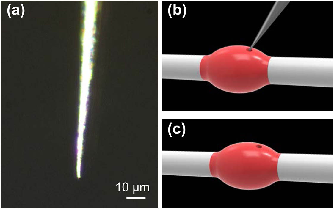
    Fig. 1. (a) Microscope image of a tungsten probe. (b), (c) Schematic illustration of surface destruction of a polymer microbottle resonator by using a tungsten probe.
    (a1)–(a3) Bright and (b1)–(b3) dark field microscope images of a microbottle resonator (Dbottle=5.3 μm, Dfiber=2.9 μm, and L=7.9 μm) and (c) their corresponding emission spectra. Electric field intensity distributions of (d) the fundamental bottle mode TM361 and (e) a typical higher-order bottle mode TM326 on the cross plane of the microresonator along its axis direction.
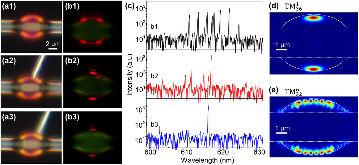
    Fig. 2. (a1)–(a3) Bright and (b1)–(b3) dark field microscope images of a microbottle resonator (Dbottle=5.3μm, Dfiber=2.9μm, and L=7.9μm) and (c) their corresponding emission spectra. Electric field intensity distributions of (d) the fundamental bottle mode TM361 and (e) a typical higher-order bottle mode TM326 on the cross plane of the microresonator along its axis direction.
    Comparison of lasing threshold between the undestroyed and destroyed microbottle resonators (Dbottle=5.1 μm, Dfiber=3.3 μm, and L=5.4 μm). Inset (a1) shows the lasing microscope image for an undestroyed microbottle and inset (b1) shows the lasing microscope image for a destroyed microbottle.
    Fig. 3. Comparison of lasing threshold between the undestroyed and destroyed microbottle resonators (Dbottle=5.1μm, Dfiber=3.3μm, and L=5.4μm). Inset (a1) shows the lasing microscope image for an undestroyed microbottle and inset (b1) shows the lasing microscope image for a destroyed microbottle.
    (a) SEM image of a small destroyed microbottle resonator (Dbottle=4.9 μm, Dfiber=3.9 μm, and L=6.1 μm). (b1), (b2) Bright and (c1), (c2) dark field microscope images and (d) their corresponding emission spectra.
    Fig. 4. (a) SEM image of a small destroyed microbottle resonator (Dbottle=4.9μm, Dfiber=3.9μm, and L=6.1μm). (b1), (b2) Bright and (c1), (c2) dark field microscope images and (d) their corresponding emission spectra.
    (a1)–(a3) Bright and (b1)–(b3) dark field microscope images and (c) their corresponding emission spectra in a microbottle resonator (Dbottle=6.1 μm, Dfiber=4.1 μm, and L=9.2 μm). (d) SEM image of the destroyed surface.
    Fig. 5. (a1)–(a3) Bright and (b1)–(b3) dark field microscope images and (c) their corresponding emission spectra in a microbottle resonator (Dbottle=6.1μm, Dfiber=4.1μm, and L=9.2μm). (d) SEM image of the destroyed surface.
    (a1)–(a3) Bright and (b1)–(b3) dark field microscope images of a microbottle resonator (Dbottle=8.1 μm, Dfiber=2.6 μm, and L=10.2 μm) and (c) their corresponding emission spectra.
    Fig. 6. (a1)–(a3) Bright and (b1)–(b3) dark field microscope images of a microbottle resonator (Dbottle=8.1μm, Dfiber=2.6μm, and L=10.2μm) and (c) their corresponding emission spectra.
    Saima Ubaid, Feng Liao, Tao Guo, Zhaoqi Gu, Shuangyi Linghu, Yanna Ma, Jiaxin Yu, Fuxing Gu. Direct single-mode lasing in polymer microbottle resonators through surface destruction[J]. Chinese Optics Letters, 2019, 17(12): 121401
    Download Citation