• Photonics Research
  • Vol. 10, Issue 3, 687 (2022)
Yufeng Gao1、2、†, Xianyuan Xia1、2、†, Lina Liu1、2, Ting Wu1、2, Tingai Chen1、2, Jia Yu1、2, Zhili Xu3, Liang Wang1、2, Fei Yan3, Zhuo Du4、5, Jun Chu1、2, Yang Zhan6, Bo Peng7, Hui Li1、2、8、*, and Wei Zheng1、2、9、*
Author Affiliations
  • 1Research Center for Biomedical Optics and Molecular Imaging, Shenzhen Key Laboratory for Molecular Imaging, Guangdong Provincial Key Laboratory of Biomedical Optical Imaging Technology, Shenzhen Institute of Advanced Technology, Chinese Academy of Sciences, Shenzhen 518055, China
  • 2CAS Key Laboratory of Health Informatics, Shenzhen Institute of Advanced Technology, Chinese Academy of Sciences, Shenzhen 518055, China
  • 3Paul C. Lauterbur Research Center for Biomedical Imaging, Institute of Biomedical and Health Engineering, Shenzhen Institute of Advanced Technology, Chinese Academy of Sciences, Shenzhen 518055, China
  • 4State Key Laboratory of Molecular Developmental Biology, Institute of Genetics and Developmental Biology, Chinese Academy of Sciences, Beijing 100101, China
  • 5Key Laboratory of Genetic Network Biology, Institute of Genetics and Developmental Biology, Chinese Academy of Sciences, Beijing 100101, China
  • 6Brain Cognition and Brain Disease Institute, Shenzhen Institute of Advanced Technology, Chinese Academy of Sciences, Shenzhen 518055, China
  • 7Centre for Micro Nano Systems and Bionic Medicine, Shenzhen Institute of Advanced Technology, Chinese Academy of Sciences, Shenzhen 518055, China
  • 8e-mail: hui.li@siat.ac.cn
  • 9e-mail: zhengwei@siat.ac.cn
  • show less
    DOI: 10.1364/PRJ.441778 Cite this Article Set citation alerts
    Yufeng Gao, Xianyuan Xia, Lina Liu, Ting Wu, Tingai Chen, Jia Yu, Zhili Xu, Liang Wang, Fei Yan, Zhuo Du, Jun Chu, Yang Zhan, Bo Peng, Hui Li, Wei Zheng. Axial gradient excitation accelerates volumetric imaging of two-photon microscopy[J]. Photonics Research, 2022, 10(3): 687 Copy Citation Text show less
    Concept and performance of Grad-TPM. (a) Grad-TPM uses a pair of gradient foci with opposite intensity distributions to successively scan the specimen and generates two images. Shallow objects appear brighter in image 1 (Im1) than in image 2 (Im2), whereas the opposite applies for deep objects. Hence, the depth of a specified object can be decoded from the intensity ratio of the two images. (b) Simplified diagram of our Grad-TPM. Phase-only spatial light modulator (SLM) was incorporated into excitation light path of Gauss-TPM to sculpt gradient foci. (c) Left panels, fluorescent beads imaged by Grad-TPM (upper) and standard Gauss-TPM (lower); right panels, colored depth 3D images of fluorescent beads obtained from two systems. Scale bar, 5 μm. (d) Absolute axial localization difference (|ze|) between Grad-TPM and Gauss-TPM of fluorescent beads. (e) Gauss-TPM depth as a function of Grad-TPM depth and statistics of the localization error (mean with standard deviation). Detailed analyses of the localization error were shown in Fig. S2 in Ref. [27]. Units of z and ze, μm.
    Fig. 1. Concept and performance of Grad-TPM. (a) Grad-TPM uses a pair of gradient foci with opposite intensity distributions to successively scan the specimen and generates two images. Shallow objects appear brighter in image 1 (Im1) than in image 2 (Im2), whereas the opposite applies for deep objects. Hence, the depth of a specified object can be decoded from the intensity ratio of the two images. (b) Simplified diagram of our Grad-TPM. Phase-only spatial light modulator (SLM) was incorporated into excitation light path of Gauss-TPM to sculpt gradient foci. (c) Left panels, fluorescent beads imaged by Grad-TPM (upper) and standard Gauss-TPM (lower); right panels, colored depth 3D images of fluorescent beads obtained from two systems. Scale bar, 5 μm. (d) Absolute axial localization difference (|ze|) between Grad-TPM and Gauss-TPM of fluorescent beads. (e) Gauss-TPM depth as a function of Grad-TPM depth and statistics of the localization error (mean with standard deviation). Detailed analyses of the localization error were shown in Fig. S2 in Ref. [27]. Units of z and ze, μm.
    Grad-TPM images of various biological structures show depth resolution and intensity contrast resembling those acquired with Gauss-TPM imaging. (a) Axons in brain slice of Thy1-GFP transgenic mouse. (b) Higher magnification views of boxed axon in (a), and depth profiles along central axis of axon. (c) Microglia in brain slice of CX3CR1-GFP transgenic mouse. First row, stitched images of two axially adjacent volumes (0–22 μm depth); second row, separate images of the upper volume (0–12 μm depth); third row, intensity images corresponding to images in the second row. (d) Depths of microglia cell bodies denoted by 1–5 in images in the first row of (c). (e) Higher magnification views of boxed microglia process in the second row of (c), and depth profiles along central axis. (f) Higher magnification views of boxed areas in the third row of (c) and corresponding intensity profiles along dashed lines for lateral resolution demonstration. Scale bars, 20 μm. Units of z, μm.
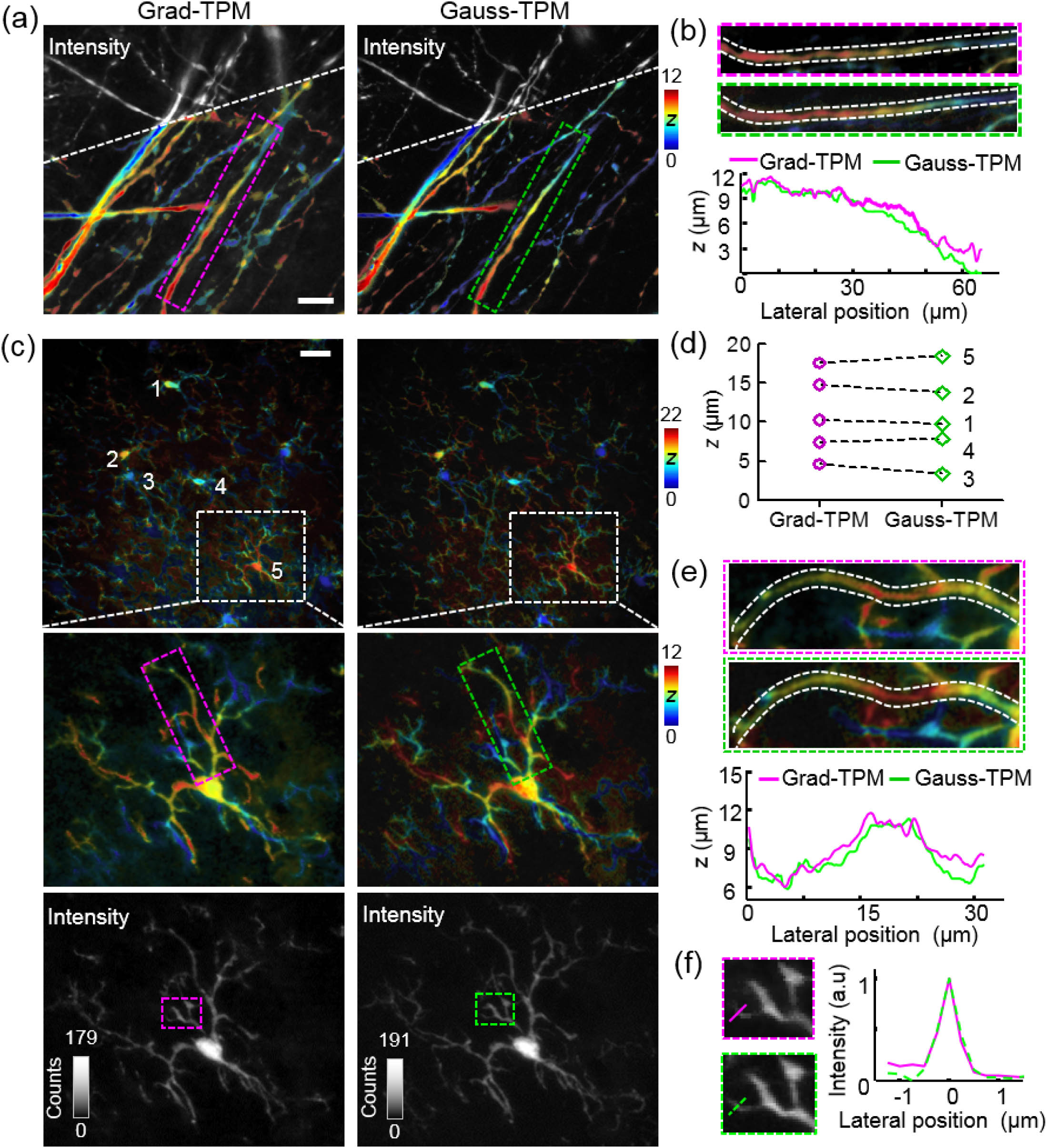
    Fig. 2. Grad-TPM images of various biological structures show depth resolution and intensity contrast resembling those acquired with Gauss-TPM imaging. (a) Axons in brain slice of Thy1-GFP transgenic mouse. (b) Higher magnification views of boxed axon in (a), and depth profiles along central axis of axon. (c) Microglia in brain slice of CX3CR1-GFP transgenic mouse. First row, stitched images of two axially adjacent volumes (0–22 μm depth); second row, separate images of the upper volume (0–12 μm depth); third row, intensity images corresponding to images in the second row. (d) Depths of microglia cell bodies denoted by 1–5 in images in the first row of (c). (e) Higher magnification views of boxed microglia process in the second row of (c), and depth profiles along central axis. (f) Higher magnification views of boxed areas in the third row of (c) and corresponding intensity profiles along dashed lines for lateral resolution demonstration. Scale bars, 20 μm. Units of z, μm.
    Grad-TPM shows observably lower photobleaching compared with traditional Gauss-TPM in (a) live cell imaging and is, therefore, highly suitable for longitudinal tracking of biological events, such as (b)–(d) phagocytosis of macrophages. (a) HEK293 cells transfected with pCAG-EGFP construct. Inset on upper right corner of each image is a magnified view of boxed area. Circled area in the inset is used to calculate average intensity to create the line graph. (b) Cultured macrophages are phagocytizing fluorescent beads. Scale bars, 20 μm. Time is shown at corner as h:min. Units of z, μm. (c) Representative trajectories of active beads (upper) and immobile beads (lower). Units of coordinate axes, μm. (d) Statistical motility parameters of immobile beads and active beads. Mean velocities indicate no significant difference (ns, two-tailed unpaired t test) of bead velocity between these two categories. Mean displacement curves suggest constrained motility in immobile beads and directed migration in active beads.
    Fig. 3. Grad-TPM shows observably lower photobleaching compared with traditional Gauss-TPM in (a) live cell imaging and is, therefore, highly suitable for longitudinal tracking of biological events, such as (b)–(d) phagocytosis of macrophages. (a) HEK293 cells transfected with pCAG-EGFP construct. Inset on upper right corner of each image is a magnified view of boxed area. Circled area in the inset is used to calculate average intensity to create the line graph. (b) Cultured macrophages are phagocytizing fluorescent beads. Scale bars, 20 μm. Time is shown at corner as h:min. Units of z, μm. (c) Representative trajectories of active beads (upper) and immobile beads (lower). Units of coordinate axes, μm. (d) Statistical motility parameters of immobile beads and active beads. Mean velocities indicate no significant difference (ns, two-tailed unpaired t test) of bead velocity between these two categories. Mean displacement curves suggest constrained motility in immobile beads and directed migration in active beads.
    Yufeng Gao, Xianyuan Xia, Lina Liu, Ting Wu, Tingai Chen, Jia Yu, Zhili Xu, Liang Wang, Fei Yan, Zhuo Du, Jun Chu, Yang Zhan, Bo Peng, Hui Li, Wei Zheng. Axial gradient excitation accelerates volumetric imaging of two-photon microscopy[J]. Photonics Research, 2022, 10(3): 687
    Download Citation