• Chinese Optics Letters
  • Vol. 15, Issue 1, 012901 (2017)
Haopeng Wu1、2, Jiulin Shi1、2、*, Feng Yan1、2, Junjie Yang1、2, Yubao Zhang1、2, and Xingdao He1、2、**
Author Affiliations
  • 1Jiangxi Engineering Laboratory for Optoelectronics Testing Technology, Nanchang Hangkong University, Nanchang 330063, China
  • 2National Engineering Laboratory for Nondestructive testing and Optoelectric Sensing Technology and Application, Nanchang Hangkong University, Nanchang 330063, China
  • show less
    DOI: 10.3788/COL201715.012901 Cite this Article Set citation alerts
    Haopeng Wu, Jiulin Shi, Feng Yan, Junjie Yang, Yubao Zhang, Xingdao He. Static light scattering properties of a ZnO nanosphere aqueous suspension at visible and near-infrared wavelengths[J]. Chinese Optics Letters, 2017, 15(1): 012901 Copy Citation Text show less
    (a) TEM image of ZnO nanospheres. (b) Transmission spectra of 1 cm pure water sample and four ZnO nanosphere/water samples. (c) Absorption spectra of 1 cm pure water sample and four ZnO nanosphere/water samples.
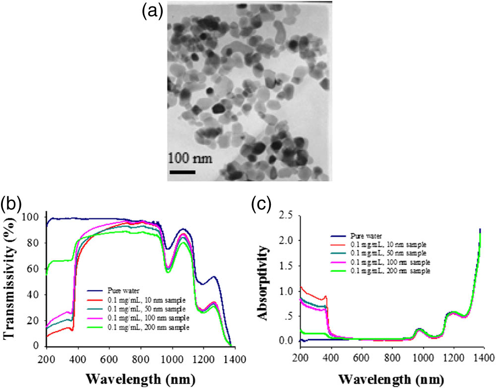
    Fig. 1. (a) TEM image of ZnO nanospheres. (b) Transmission spectra of 1 cm pure water sample and four ZnO nanosphere/water samples. (c) Absorption spectra of 1 cm pure water sample and four ZnO nanosphere/water samples.
    Mie scattering in ZnO sample and Rayleigh scattering in pure water at three different incident wavelengths. (a) Incident wavelength 457 nm. (b) Incident wavelength 532 nm. (c) Incident wavelength 633 nm.
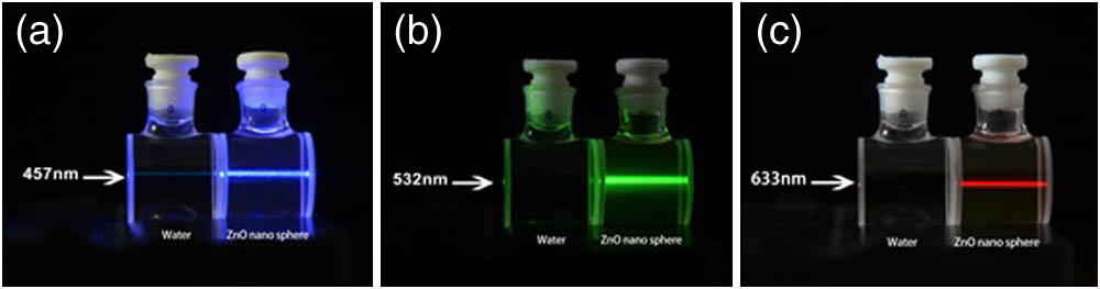
    Fig. 2. Mie scattering in ZnO sample and Rayleigh scattering in pure water at three different incident wavelengths. (a) Incident wavelength 457 nm. (b) Incident wavelength 532 nm. (c) Incident wavelength 633 nm.
    Experimental setup for measuring the angular distribution of the scattering intensity.
    Fig. 3. Experimental setup for measuring the angular distribution of the scattering intensity.
    Measured normalized angular distribution curves for four scattering samples with different particle diameters. The blue arrow denotes the propagation direction of the incident laser.
    Fig. 4. Measured normalized angular distribution curves for four scattering samples with different particle diameters. The blue arrow denotes the propagation direction of the incident laser.
    Calculated normalized angular distribution of scattering intensity of four scattering samples with different particle diameters. The long dashed lines, dashed–dotted lines, and solid lines represent that the polarization of the incident light is vertical linear polarization (I1), parallel linear polarization (I2), and 45° linear polarization (or naturally polarized light, I1+I2/2), respectively.
    Fig. 5. Calculated normalized angular distribution of scattering intensity of four scattering samples with different particle diameters. The long dashed lines, dashed–dotted lines, and solid lines represent that the polarization of the incident light is vertical linear polarization (I1), parallel linear polarization (I2), and 45° linear polarization (or naturally polarized light, I1+I2/2), respectively.
    3D plot of the normalized angular distribution of scattering intensity versus the scattering angle and the particle size. (a) 3D line plot. (b) 3D mesh plot. The direction of the 0° or 360° scattering angle represents the propagation direction of the incident laser.
    Fig. 6. 3D plot of the normalized angular distribution of scattering intensity versus the scattering angle and the particle size. (a) 3D line plot. (b) 3D mesh plot. The direction of the 0° or 360° scattering angle represents the propagation direction of the incident laser.
    Photos for observing the change of forward scattering of four samples with different particle diameters. The wavelength of the incident laser is 457 nm. The horizontal yellow arrows represent the direction of the incident laser.
    Fig. 7. Photos for observing the change of forward scattering of four samples with different particle diameters. The wavelength of the incident laser is 457 nm. The horizontal yellow arrows represent the direction of the incident laser.
    Scattering extinction coefficients of four different ZnO samples.
    Fig. 8. Scattering extinction coefficients of four different ZnO samples.
    Experimental and theoretical curves of scattering cross sections of four different ZnO samples versus the wavelength. The corresponding particle diameters are (a) 10, (b) 50, (c) 100, and (d) 200 nm.
    Fig. 9. Experimental and theoretical curves of scattering cross sections of four different ZnO samples versus the wavelength. The corresponding particle diameters are (a) 10, (b) 50, (c) 100, and (d) 200 nm.
    Scattering extinction sections of four ZnO samples at the three given wavelengths.
    Fig. 10. Scattering extinction sections of four ZnO samples at the three given wavelengths.
    Haopeng Wu, Jiulin Shi, Feng Yan, Junjie Yang, Yubao Zhang, Xingdao He. Static light scattering properties of a ZnO nanosphere aqueous suspension at visible and near-infrared wavelengths[J]. Chinese Optics Letters, 2017, 15(1): 012901
    Download Citation