
Song Siyu, Li Zhongliang, Gao Yunhua, Yu Junjie, Nan Nan, Wang Xuan, Yuan Chunxiao, Wang Xiangzhao. Swept Source Optical Coherence Tomography System for Transdermal Drug Delivery Imaging by Microneedles[J]. Chinese Journal of Lasers, 2018, 45(8): 807001
- Chinese Journal of Lasers
- Vol. 45, Issue 8, 807001 (2018)
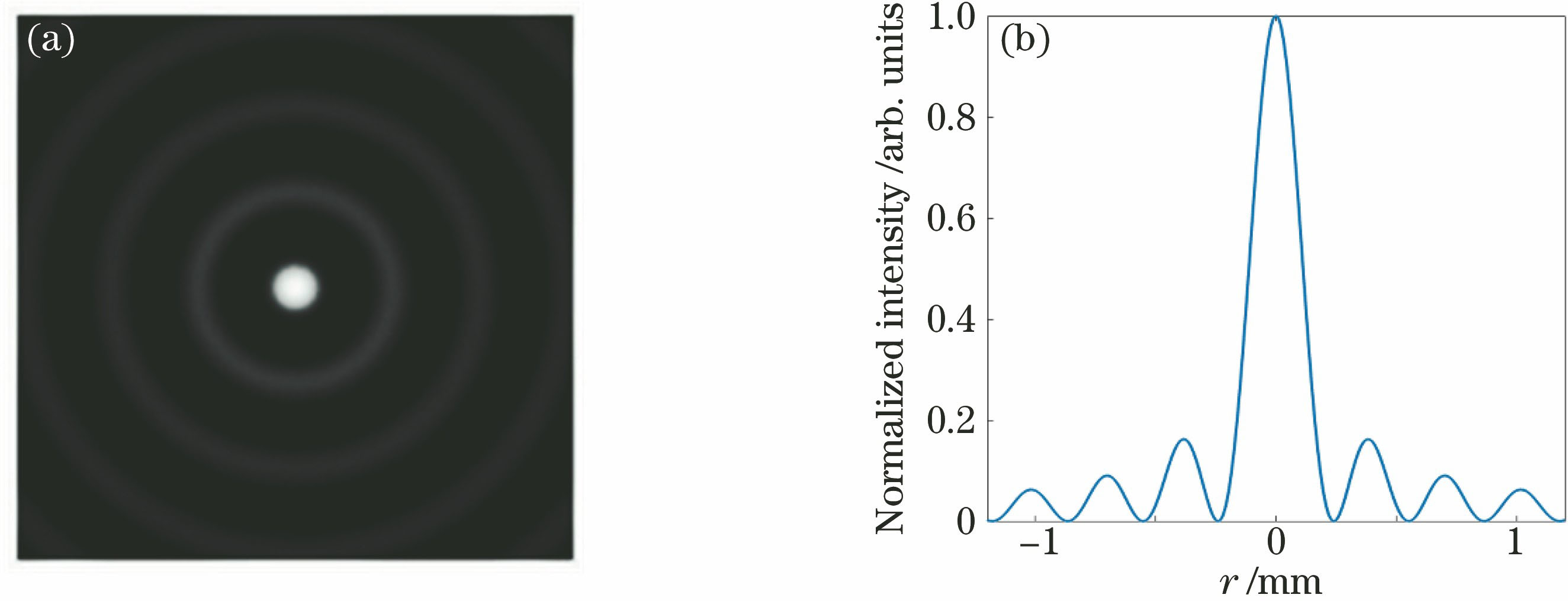
Fig. 1. (a) Intensity distribution in cross-section; (b) one-dimensional horizontal intensity distribution

Fig. 2. Schematic of high-quality Bessel beam generated by CDG
Fig. 3. Schematic of SSOCT system (the solid line represents the optical signal and the dotted line represents the electrical signal)
Fig. 4. Flow chart of data processing algorithm
Fig. 5. (a) Axial point spread function; (b) image of 3 μm microbeads; (c)-(e) lateral point spread functions
Fig. 6. Schematic of a microneedle array and its geometrical shape
Fig. 7. (a) Image of microneedle array inserted into the skin; (b) OCT image before inserting the microneedle; (c)-(e) OCT images of microneedle array inserted into the skin for 0, 30, 60 min, respectively; (f) OCT image after removing the microneedle base plate

Set citation alerts for the article
Please enter your email address
© Copyright 2018-2021 | Chinese Laser Press. All Rights Reserved 沪ICP备15018463号-20