
J. Qian, G. D. Wang, K. Y. Lou, D. Y. Shen, Q. Fu, Q. Z. Zhao. Self-induced birefringence of white-light continuum generated by interaction of focused femtosecond laser pulses with fused silica[J]. High Power Laser Science and Engineering, 2020, 8(2): 02000e19
- High Power Laser Science and Engineering
- Vol. 8, Issue 2, 02000e19 (2020)
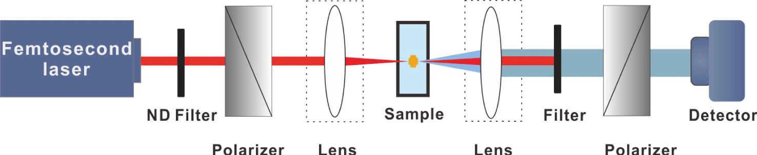
Fig. 1. Schematic diagram of self-induced birefringence of white-light continuum.
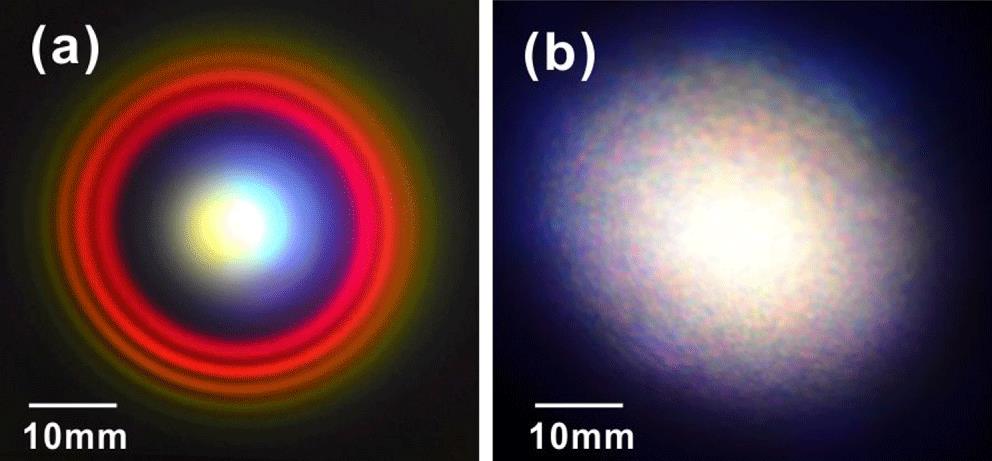
Fig. 2. Original color photographic image of continuum taken at a distance of 180 mm from the focus center when the laser pulse energy is (a) 10 μJ and (b) 550 μJ.
Fig. 3. Beam profile evolution of the transmitted light signal with varied pulse energy and laser exposure time.
Fig. 4. Time evolution of the transmitted signal of the generated continuum at varied pulse energy. Inset of (a): saturation value of the transmitted signal as a function of the pump energy.
Fig. 5. Optical images of the femtosecond laser induced structure under the illumination of light on a transmission microscope with (a) parallel polarizer and (b) crossed polarizer on both sides of the samples. Time evolution of the birefringence structure under cross-polarization illumination at pulse energy of (c) 90 μJ and (d) 550 μJ.  represents the laser propagation direction. The red dashed line indicates the focal depth.
represents the laser propagation direction. The red dashed line indicates the focal depth. Fig. 6. The dependence of the transmitted continuum behind the second polarizer (a) on the laser exposure time at pulse energy of 550 μJ and (b) on the pulse energy at laser exposure time of 400 s.

Set citation alerts for the article
Please enter your email address
© Copyright 2018-2021 | Chinese Laser Press. All Rights Reserved 沪ICP备15018463号-20