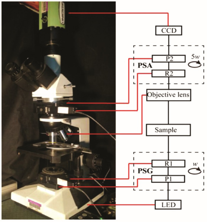
Significance Polarization imaging technology has the advantages of non-invasive detection, rich information, sensitivity to the microstructure of the sample, and compatibility with traditional optical imaging technology, which makes it suitable for combining with microscopy techonology based on staining methods to distinguish the characteristics of different microstructures of pathological tissues. By adding the polarization state analyzer (PSA) and polarization state generator (PSG) modules to the commercial transmission and colinear reflection optical microscopes, the Mueller matrix microscopic imaging can be performed. The Mueller matrix can realize the complete characterization of the polarization properties of the sample.
Progress We have established the upright transmission Mueller matrix microscope (
In this article, we also introduce some biomedical applications of the Mueller matrix microscope. By perfoming Mueller matrix imaging of cancerous liver tissues in different stages and calculating polarization parameters, it can realize the characterization of the degree of liver fibrosis in the cancerous liver tissue (Figs. 5--7). By combining with data technologies including machine learning, new polarization parameters used to quantitatively characterize the microstructure of biological tissues can be derived, which can accurately distinguish specific pathological structures (
Conclusions and Prospects In this article, we summarize several modular polarization microscopes implemented in our previous studies, including the transmission Mueller matrix microscope and the collinear reflection Mueller matrix microscope based on dual rotating retarders, as well as the transmission Mueller matrix microscope based on dual linear polarization CCDs. Then we introduce some applications of modular polarization microscope in the biomedicine field. With the combination of microscopy and polarization imaging technology, Mueller matrix microscope can be directly upgraded from ordinary optical microscopy methods. It has the following advantages: suitable for the studies of biological living systems, capable of obtaining cross-scale image information, and easy to be combined with data science technologies. Besides biomedicine, the Mueller matrix microscope can be also applied to many fields including material science, defect detection, etc. There also exsit some aspects that need to be improved: relatively small imaging area, and high requirement for system stability and residual polarization artifacts of the optics inside the system. This article puts forward specific suggestions for the above problems. In the future, the development tread of Mueller matrix microscope is faster measurement speed and higher measurement accuracy. With the improvements of polarization modulation and measurement technology, the Mueller matrix microscope is expected to perform real-time and accurate full-polarization measurement of living cells and in-vivo tissues, and becomes an important tool to promote biomedical applications.