
Huifang Liu, Dongqin Lei, Fen Qin, Sijia Wang, Zhenxi Zhang. Multi-Responsive Liposomal Nanocomplex Encapsulating Ce6 & MMP-2 Inhibitors for Photodynamic-Immune Synergistic Treatment of Melanoma[J]. Chinese Journal of Lasers, 2023, 50(3): 0307203
- Chinese Journal of Lasers
- Vol. 50, Issue 3, 0307203 (2023)
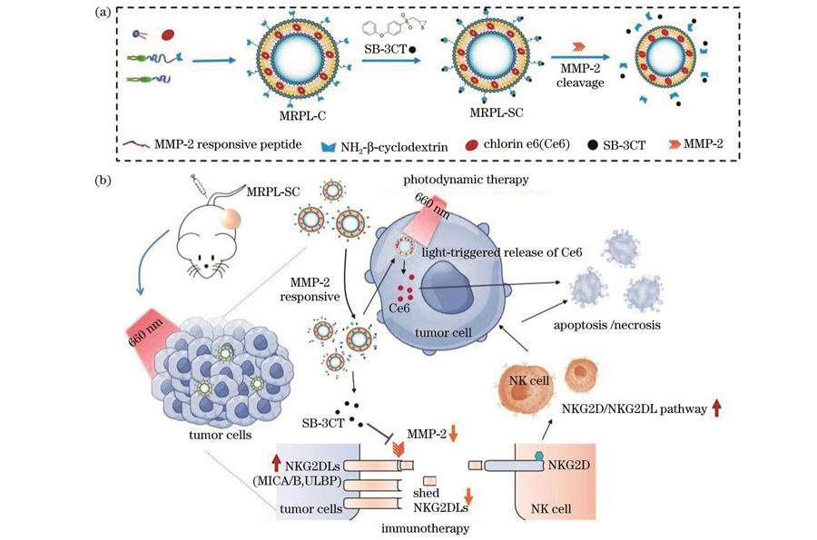
Fig. 1. Schematic diagrams of MRPL-SC liposomes. (a) MRPL-SC synthesis and MMP-2 responsive release; (b) dual-responsive drug release and mechanisms to improve photodynamic immunotherapy of MRPL-SC in tumor
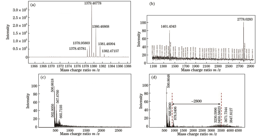
Fig. 2. MALDI-TOF spectrometry detection results. (a) MRP; (b) MRP-β-CD; (c) DSPE-PEG(3400)-Mal; (d) DSPE-PEG(3400)-MRP-β-CD
Fig. 3. Characterization of MRPL-SC. (a) Synthesis schematic of MRPL-SC; (b) particle size distributions and Cryo-TEM characterization of MRPL-C and MRPL-SC loaded with SB-3CT; (c) UV-vis spectra of free SB-CT, Ce6 and liposomes, where blank loads no drug, MRPL-C loads only Ce6, MRPL-S loads only SB-3CT, and MRPL-SC loads both Ce6 and SB-3CT; (d) singlet oxygen generation
Fig. 4. MMP-2 responsiveness of MRPL. (a) Drug release schematic illustration of MRPL-SC with MMP-2 responsive; (b) particle size distributions and Cryo-TEM images of MRPL-SC incubated with rhMMP-2; (c) SB-3CT release curves of MRPL-SC incubated with different mass concentrations of MMP-2; (d) Ce6 release before and after laser triggering
Fig. 5. Cellular uptake and cytotoxicity of MRPLs in vitro. (a) Fluorescent images of Ce6 uptake by A375 cells (scale bar: 10 μm); (b) relative quantitative analysis of fluorescence intensity of Ce6 uptake by A375 cells; (c) fluorescent images of ROS produced by A375 cells after different treatments; (d) cell viability of A375 cells treated by MRPL without laser irradiation; (e) cell viability of A375 cells treated by MRPL with laser irradiation
Fig. 6. Mechanism of NKG2D/NKG2DL pathway in A375 cells treated with MRPLs. (a) Protein expression of NKG2D/NKG2DL pathway after MRPLs treatment with or without laser; (b) soluble MICA and MICB shedding from A375 cells treated with MRPLs after 48 h incubation; (c) proposed signaling pathway of MRPL-SC upregulating the expression of NKG2DLs on the surface of A375 cancer cells; (d) killing effect of NK cells at different effector-target ratios (scale bar: 100 μm); (e) NK cell-mediated cytotoxicity to A375 cells at effector-target ratio of 4∶1
Fig. 7. In vivo fluorescence imaging and biodistribution analysis of nude mice bearing tumors after tail vein injection of Ce6 and MRPL-C. (a) Schematic diagram of tumor-bearing mouse imaging; (b) in vivo fluorescence imaging of A375-bearing mice after intravenous injection of Ce6 and MRPL-C nanoparticles at different times; (c) fluorescence intensity of major organs and tumor dissection 24 h after intravenous injection
Fig. 8. In vivo anti-tumor effect of MRPL-SC (**P < 0.01, ***P < 0.001). (a) Schematic illustration of in vivo therapeutic time line on tumor-bearing mouse; (b) tumor volume change of mice (n=6) from various treatment groups during 14 d treatment; (c) relative tumor proliferation rate of tumor-bearing mouse in different treatment groups; (d) tumor weight of tumor in different treatment groups; (e) H&E and TUNEL staining of tumor sections of different treatment groups excised two days after irradiation
Fig. 9. H&E staining results of major organs of tumor-bearing mice for different treatments (scale bar: 50 μm)

Set citation alerts for the article
Please enter your email address
© Copyright 2018-2021 | Chinese Laser Press. All Rights Reserved 沪ICP备15018463号-20