
Hong Jia, Hongming Jiang, Yuping Zhang, Shuxu Hua, Qing Liu, Yuquan Yuan, Yanfei Hu, Feng Peng, Xiaofeng Liu. Enhanced near-infrared light-induced photoresponse via transition of monocrystalline phase and surface reconstruction[J]. Chinese Optics Letters, 2023, 21(5): 051603
- Chinese Optics Letters
- Vol. 21, Issue 5, 051603 (2023)
Abstract
Keywords
1. Introduction
Upconversion (UC) luminescent materials have been extensively used in display imaging, sensing detection, photothermal therapy, and photovoltaics because of their peculiar optical process[1–7]. Metal ions with the d, f orbitals can fulfill the requirement for the UC by virtue of their long-lived excited states and ladder-level configurations[8]. In addition, a large number of transition metals and actinide-doped materials have been reported to display UC properties[9]. Among them, lanthanide-doped UC solid materials show superior upconversion luminescence (UCL) characteristics, and fluoride and sulfide UC materials with low phonon energies are the ideal host matrix[10,11]. However, the matrix is being replaced by oxide materials due to its poor stability and high toxicity. In oxide UC materials, the inherent structural defects, low absorption cross section, and dipole transitions of the rare-earth ions usually result in a low UC efficiency. Up to the present, several strategies have been developed to optimize the materials design and to improve the efficiency of oxide UC materials.
In order to reduce the quenching effect of surface defects or surface-related ligands, a core-shell structure is one of the solutions. For example, Vetrone et al. reported the use of “active” doped shells to increase surface passivation to reduce nonradiative transition rates and to enhance UCL[12]. Liu et al. also achieved a significant enhancement of the UCL by adding a microlens array to the top of the UC nanomaterial to modulate the temporal distribution of transmitted excitation photons[13,14]. However, the UC process is still relatively inefficient due to the inherently low absorption coefficients of rare-earth (RE) ions. This problem can be mitigated by using dye sensitization for RE ions, and the combination of dye sensitization with a core-shell structure could lead to higher efficiency. For instance, Shao and coworkers introduced the organic dye indocyanine green (ICG) onto the surface of core-shell structured -based UC particles[15], which demonstrated a large increment in UC emission intensity several times and enabled a wider excitation band. However, organic dyes are not suitable for long-term solar illumination due to their poor molecular stability and susceptibility to competitive energy loss during heterogeneous energy transfer. In atomic spectroscopy, electric dipole transition is one of the most important transitions in the process of the atom/nucleus emission or absorption of photons[16]. The lack of an electric dipole moment will result in prohibition of transition. Photonic crystal (Phc) structure engineering by incorporation of upconverting nanoparticles (UCNPs) into a photonic lattice could boost the UC intensity due to the enhanced local electric field under NIR excitation[17]. A similar strategy for electric field enhancement based on plasmonic structures has also been found to be highly effective in enhancing the UC emission[18–23], but the use of precious metals and the high fabrication cost make it difficult for larger-scale applications.
In the above UCL enhancement mechanisms, local field modulation and surface optimization are always the most efficient strategies. Due to the influence of many restrictive conditions on the photovoltaic conversion, we here chose surface engineering strategy by using non-RE ions doping to adjust the atomic structure of the UCNPs’ surface and to control structure transition from amorphous to ordered crystal to increase the absorption efficiency and enhance the luminescence efficiency. Due to the low phonon energy, excellent thermal stability, high physical durability, and the close cation ionic radius, is considered as a good host material for rare-earth ions[24,25]. ions with ladder energy levels and long excited state life are considered the most suitable UC activator[26–28]. In addition, we consider that the local fields and surface microstructure may be changed by doping with alkali metal ions due to the differences in ionic radii and the electronegativity. Although most previous work employed ions as the co-dopant[29,30], we show that , X (, , ) exhibits large enhancements in UC emission compared with the phosphor without the introduction of alkali metal elements. In addition, we propose a low-cost strategy that rationally integrates the UC particles with Si-based photoresistors (Si-BPRs) for NIR responsive devices. With the conversion of the NIR () photons into visible light (400 to 700 nm) by UC materials, the developed device demonstrates strong electrical responses to NIR light, suggesting its potential use in NIR sensing and detection.
Sign up for Chinese Optics Letters TOC. Get the latest issue of Chinese Optics Letters delivered right to you!Sign up now
2. Experiment Section
3. Results and Discussion
3.1. Structural and morphological characteristics
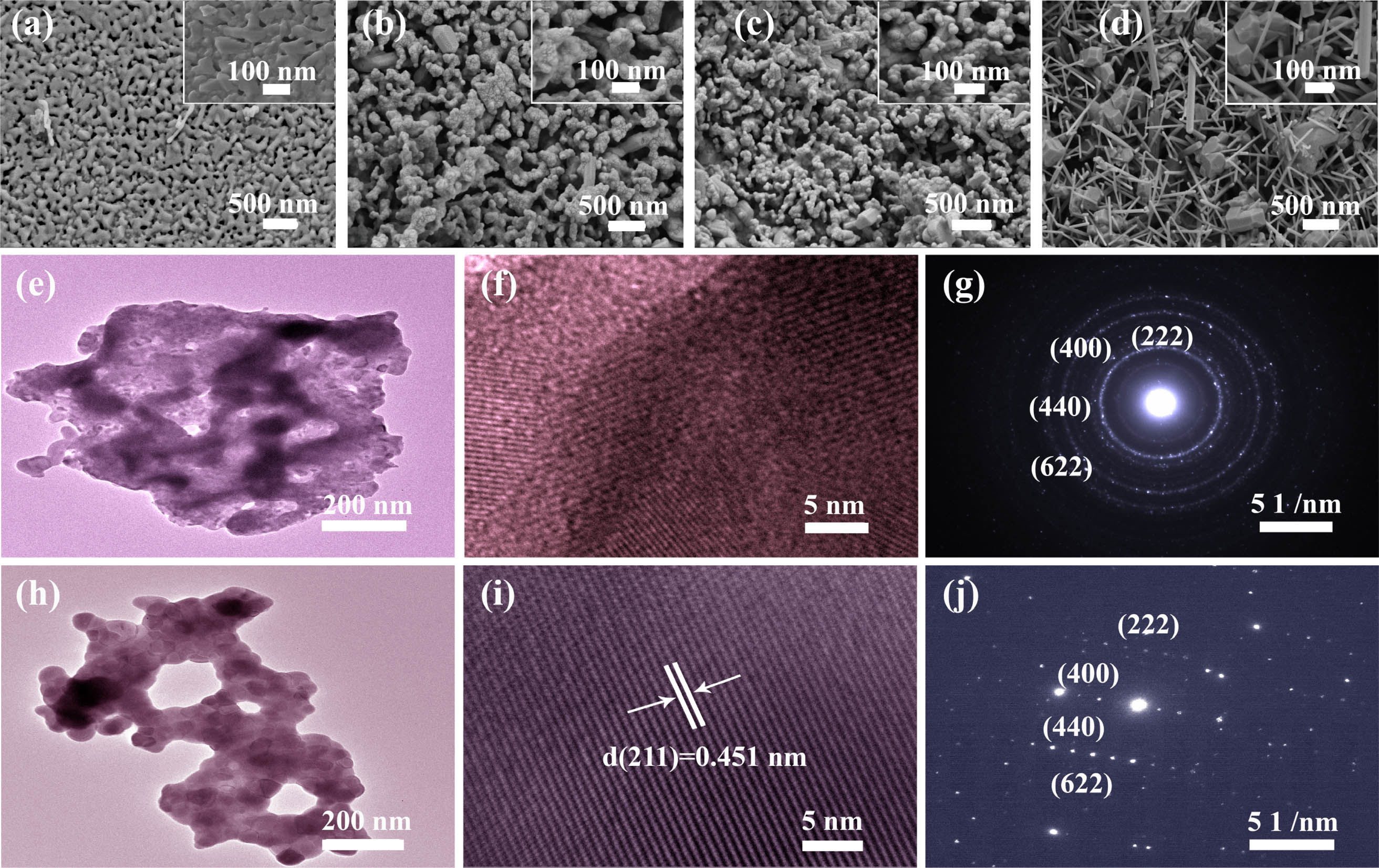
In order to observe the morphology of changes of its morphology and structure after the introduction of alkali metals, the and , (M = Na, K, Cs) phosphors were recorded by an SEM. As shown in Fig. 1(a), the phosphor particles exhibit a tabular structure with a radius of 80–300 nm. After the introduction of and ions, the tabular structure was significantly changed. Figures 1(b) and 1(c) show that the size of nanoparticles doped with and ions is 60–100 nm and 60–250 nm, respectively. In addition, the introduction of a large number of with large ionic radii leads to the formation of bar (30–130 nm) and columnar (230–450 nm) morphologies. The possible mechanism of morphology evolution after introducing ions into phosphor is proposed as follows. The ions likely enter into interstitial sites due to their different valence states with ions when is introduced into the matrix. Similar to the vast number of ions doping, this will inevitably cause some lattice distortion[31–33]. However, doping a large number of ions with large ionic radii may cause a greater degree of lattice distortion and even restrict the periodic arrangement of some crystal planes. The high chemical potential energy at high temperatures results in the rapid aggregation of primary particles. While the continuous self-assembly of easily grown crystal faces in the process of mutual reactions results in long columnar and strip-like forms with the same lattice orientation [Fig. 1(d), and Fig. S3(d) in the Supplementary Material][34], the analysis of , 10% phosphors by TEM, high-resolution transmission electron microscopy (HRTEM), and SAED is helpful for further observation of their morphological characteristics. As shown in Figs. 1(e) and 1(h) transmission microscopic images, a phosphor is formed by the polymerization of particles with a diameter of about 20–40 nm. The introduction of ions greatly reduced the phosphor agglomeration phenomenon and increased the size of nanoparticles to about 60 nm, which is similar to the results obtained in Table S1 (Supplementary Material). On the other hand, the transmission amplification results in Fig. 1(e) indicate that the 80–300 nm tabular structure in Fig. 1(a) may be composed of smaller particles with a diameter of about 20–40 nm, while the introduction of alkali metals is conducive to reducing the agglomeration phenomenon. Meantime, HRTEM showed different lattice fringes [Figs. 1(f) and 1(i)]. Compared with , the introduction of ions contributes to the formation of the monocrystalline phase, and the interplanar spacing is about 4.51 Å (211 planes). This is undoubtedly beneficial for many aspects, such as eliminating high dislocation defects or high concentrations of impurities. In addition, the SAED patterns given in Figs. 1(g) and 1(j) can be indexed to the (222), (400), (440), and (622) planes of cubic . Therefore, for and , 10% phosphors, although the doping of ions caused a large change in particle morphology, it only leads to a small change in the phase. In addition, the morphologic features of - and -doped are shown in Fig. S3 (see Supplementary Material).
![]()
Figure 1.SEM images of the synthesized samples: (a) Gd2O3: 3% Er3+; (b) Gd2O3: 3% Er3+, 10% Na+; (c) Gd2O3: 3% Er3+, 7% K+; (d) Gd2O3: 3% Er3+, 3% Cs+. (e), (h) TEM images; (f), (i) HRTEM images; (g), (j) SAED patterns of Gd2O3: 3% Er3+ and Gd2O3: 3% Er3+, 10% Na+ phosphors.
3.2. Optical performance
The absorption spectra and the emission spectra of the samples prepared under different conditions were studied. Figure 2(a) presents the absorption spectra for and , (M = Na, K, Cs; y = 10%, 7%, 3%). All samples showed the expected absorption peaks at 808 and 980 nm. For the absorption peak at 980 nm, it was found that the absorption peak was enhanced when ions were added, but the introduction of and ions had no observable effect. In addition, the UC photoluminescence (PL) spectra of the samples are greatly affected by the doping of different concentrations of active ions. Therefore, the PL emission spectra of the prepared samples were studied in more detail. Figure 2(b) shows the emission spectra of (x = 1%, 3%, 5%, 7%, 10%, 12%) phosphors excited by a 980 nm laser. All the PL spectra showed the same spectral shape and reached the maximum peak at the green region (539–570 nm). When the concentration exceeds 3%, the PL intensity decreases notably as a result of concentration quenching. It is also possible that the solubility limit of in the lattice is smaller considering the mismatch in ionic size. In order to explore the effect of alkali metal ions (, , ) on the luminescence of phosphors, the PL emission spectra of corresponding phosphors were studied. For , phosphors in Fig. 2(c), the peak position does not move with the addition of ions. This indicates that the introduction of ions can significantly increase the intensity of the emission peak in the phosphors, but the local structure of remains unchanged. The luminescence is strongest when the concentration of ions reaches 10%. Similarly, the optimal concentrations of and ions were 7% and 3%, respectively [Fig. 2(d)], and the emission intensities were ordered as . The spectral result in Fig. 2(d) shows that the strongest peak is located at 563 nm. In addition, the left image of Fig. 2(d) shows the visible green luminescence of the samples at 980 nm excitation. The results shown in the green luminescence diagram are fully synchronized with the curve in Fig. 2(d), which further proves that the addition of alkali metals () can significantly improve the luminescence intensity.
![]()
Figure 2.(a) Absorption spectra of Gd2O3: 3% Er3+ and Gd2O3: 3% Er3+, yM+ (M = Na, K, Cs; y = 10%, 7%, 3%). UC PL emission spectra for (b) Gd2O3: xEr3+ (x = 1%, 3%, 5%, 7%, 10%, 12%); (c) Gd2O3: 3% Er3+, yNa+ (y = 1%, 3%, 5%, 7%, 10%, 12%); and (d) Gd2O3: 3% Er3+, zM+ (M = Na, K, Cs; z = 10%, 7%, 3%). (e) Luminescence intensity of the Gd2O3: 3% Er3+, 10% Na+ phosphors at different annealing temperatures; (f) CIE chromaticity diagram of the Gd2O3: 3% Er3+ and the Gd2O3: 3% Er3+, 10% Na+ phosphors.
Here, we also recorded the luminescence intensity of , M (, , and ) samples after 60 days at room temperature. As shown in Fig. S3(a) (see Supplementary Material), phosphors show a large intensity attenuation with the increase of storage time. There is no doubt that this is closely related to prolonged exposure to air. The possible reason is that the ions could interact with water molecules in air, which results in the weakening of the luminescence intensity with time. Fortunately, the emission intensity of phosphors before storage is comparable to that of -doped phosphors [Fig. 2(a)]. There are two reasons for this. (1) The ion has the strongest metal properties, the position of lowest unoccupied molecular orbital (LUMO) level increases and the forbidden bandwidth becomes narrower when it enters into the matrix, leading to the enhancement of the transition probability from highest occupied molecular orbital (HOMO) level to the conduction band. (2) The introduction of ions optimized the surface properties of the phosphor. Compared with the polycrystalline phase for the ion-doped samples [Figs. S3(b) and S3(c) in the Supplementary Material], the phosphor forms a single crystal phase that is beneficial to generate efficient PL [Figs. S3(e) and S3(f) in the Supplementary Material] because compared with the polycrystalline phase, the monocrystalline phase is more favorable to the light refraction and scattering process. However, the vacuum environment becomes a necessary condition for the practical application of phosphors considering the high sensitivity to air. Figure 2(e) shows PL emission spectra as a function of annealing temperature. Obviously, with the increase in annealing temperature, the luminescence intensity increases continuously and reaches its maximum at 1100°C. The luminescence images in the inset of Fig. 2(e) again substantiate the obvious enhancement of luminescence intensity at 1100°C. To visualize the emission of the phosphor, the Commission International de I’Eclairage (CIE) 1931 chromaticity coordinates pattern of the and samples are calculated. As shown in Fig. 2(f), the CIE coordinates of and phosphors are calculated as , and , , which fit well in the green areas.
The UCL mechanism of , co-doped phosphors under 980 nm laser excitation was investigated. It is well known that the spectral intensity () of UC has a nonlinear relationship with the excitation power density (). For the unsaturated UC process, the number of photons required to transition from one level to another satisfies the following formula[35,36]:
![]()
Figure 3.(a), (b) Logarithmic patterns of power dependence of 3% Er3+ and 3% Er3+, 10% Na+-doped Gd2O3 sample materials in red and green wavebands; (c) emission attenuation curves of Gd2O3: 3% Er3+ and Gd2O3: 3% Er3+, 10% Na+ phosphors were fitted by a double exponential decay function (980 nm excitation); (d) possible UC mechanism of Er3+ ions under 980 nm excitation.
The UC phosphor particles were then integrated with a Si photoresistor for the demonstration of NIR photoresponse. In this device (see Section 2), the photocurrent response to the NIR light is related to the UC emission of the phosphor particles. In this work, the photocurrent responses of devices based on the and doped phosphors that were excited by the concentrated NIR part of sunlight (by using a filter) were studied. In our experiment, a transparent gel formed by mixing phosphor powder and polymer is coated on the Si photoresistor to realize the conversion of the NIR light to an electrical response signal. Here, the excitation light is mechanically chopped to form a square-shaped signal with 2 s duration. The device shows no photovoltage response in a dark environment. Under optical excitation, the device based on the composite film only shows a weak photovoltage response of about 0.2 V, as shown in Fig. 4. In contrast, the photovoltage response of the device based on composite film increased to 1.4 V. The results clearly suggest that composite film has a higher UC efficiency, so that the light response to the NIR part of the sunlight can be greatly enhanced. In addition, our device is stabler due to the encapsulation effect of the phosphor coated by a transparent polymer[37,38]. These results provide an effective strategy for the design and application of NIR light-responsive UC optoelectronic devices.
![]()
Figure 4.Photocurrent response of the device incorporating Gd2O3: 3% Er3+/PMMA and Gd2O3: 3% Er3+, 10% Na+/PMMA composite films under the square pulse with an NIR wavelength of 980 nm, the applied potential of 0 V, and 2 s off/on cycles.
4. Conclusions
In summary, the crystal structure and surface properties of and , (M = Na, K, Cs) phosphors have been systematically studied by a large number of microscopic methods. We are committed to discovering the universal features of enhanced UC efficiency after the introduction of alkali metal ions. The results show that the monocrystalline phase transition and surface optimization induced by the introduction of , , and ions in phosphors can significantly improve the UC intensity of phosphors. Meantime, the bar and column morphology may be more conducive to the UC PL process. In addition, an NIR-responsive device was demonstrated by coating a Si photoresistor with a transparent gel incorporating the UC powders. The photoelectric voltage is only 0.2 V for the device based on the film, while it goes up to 1.4 V for the device using film. It is worth noting that, although ions doping has a higher UC intensity, it faces a large instability. In comparison, ions have a higher performance due to their ubiquity and high UC efficiency.
References
[1] T. Han, Y. Wang, S. Ma, M. Li, N. Zhu, S. Tao, J. Xu, B. Sun, Y. Jia, Y. Zhang, S. Zhu, B. Yang. Near-infrared carbonized polymer dots for NIR-II bioimaging. Adv. Sci., 9, 2203474(2022).
[2] M. Yang, J. Huang, J. Fan, J. Du, K. Pu, X. Peng. Chemiluminescence for bioimaging and therapeutics: recent advances and challenges. Chem. Soc. Rev., 49, 6800(2020).
[3] H. Huang, S. Wang, R. Chen, N. Zhang, H.-R. Yao, Y. Zheng, F. Huang, D. Chen. Engineering upconverting core-shell nano-probe for spectral responsive fluid velocimetry. Nano Res., 16, 1212(2022).
[4] M.-P. Zhuo, X.-D. Wang, L.-S. Liao. Recent progress of novel organic near-infrared-emitting materials. Small Sci., 2, 2200029(2022).
[5] H. Altug, S. H. Oh, S. A. Maier, J. Homola. Advances and applications of nanophotonic biosensors. Nat. Nanotechnol., 17, 5(2022).
[6] J. Li, L. Liu, X. Chen, C. Lu, H. Zhang, B. Li, C. Xue, Z. Lou. Dual-functional nonmetallic plasmonic hybrids with three-order enhanced upconversion emission and photothermal bio-therapy. Laser Photonics Rev., 16, 2200197(2022).
[7] F. Zhang, S. Y. Park, C. Yao, H. Lu, S. P. Dunfield, C. Xiao, S. Uličná, X. Zhao, L. D. Hill, X. Chen, X. Wang, L. E. Mundt, K. H. Stone, L. T. Schelhas, G. Teeter, S. Parkin, E. L. Ratcliff, Y.-L. Loo, J. J. Berry, M. C. Beard, Y. Yan, B. W. Larson, K. Zhu. Metastable Dion-Jacobson 2D structure enables efficient and stable perovskite solar cells. Science, 375, 71(2022).
[8] L. Yang, J. Luo, L. Gao, B. Song, J. Tang. Inorganic lanthanide compounds with f-d transition: from materials to electroluminescence devices. J. Phys. Chem. Lett., 13, 4365(2022).
[9] X. Feng, L. Lin, R. Duan, J. Qiu, S. Zhou. Transition metal ion activated near-infrared luminescent materials. Prog. Mater. Sci., 129, 100973(2022).
[10] A. Shalav, B. S. Richards, T. Trupke, K. W. Krämer, H. U. Güdel. Application of NaYF4:Er3+ up-converting phosphors for enhanced near-infrared silicon solar cell response. Appl. Phys. Lett., 86, 013505(2005).
[11] W. Chen, A. G. Joly, J. Z. Zhang. Up-conversion luminescence of Mn2+ in ZnS:Mn2+ nanoparticles. Phys. Rev. B, 64, 041202(2001).
[12] F. Vetrone, R. Naccache, V. Mahalingam, C. G. Morgan, J. A. Capobianco. The active-core/active-shell approach: a strategy to enhance the upconversion luminescence in lanthanide-doped nanoparticles. Adv. Funct. Mater., 19, 2924(2009).
[13] Q. Liu, H. Liu, D. Li, W. Qiao, G. Chen, H. Ågren. Microlens array enhanced upconversion luminescence at low excitation irradiance. Nanoscale, 11, 14070(2019).
[14] Y. Ji, W. Xu, N. Ding, H. Yang, H. Song, Q. Liu, H. Agren, J. Widengren, H. Liu. Huge upconversion luminescence enhancement by a cascade optical field modulation strategy facilitating selective multispectral narrow-band near-infrared photodetection. Light Sci. Appl., 9, 2047(2020).
[15] W. Shao, G. Chen, A. N. Kuzmin, H. L. Kutscher, A. Pliss, T. Y. Ohulchanskyy, P. N. Prasad. Tunable narrow band emissions from dye-sensitized core/shell/shell nanocrystals in the second near-infrared biological window. J. Am. Chem. Soc., 138, 16192(2016).
[16] H. Dong, L. D. Sun, C. H. Yan. Basic understanding of the lanthanide related upconversion emissions. Nanoscale, 5, 5703(2013).
[17] C. Mao, K. Min, K. Bae, S. Cho, T. Xu, H. Jeon, W. Park. Enhanced upconversion luminescence by two-dimensional photonic crystal structure. ACS Photonics, 6, 1882(2019).
[18] J. Xu, Z. Dong, M. Asbahi, Y. Wu, H. Wang, L. Liang, R. J. H. Ng, H. Liu, R. A. L. Vallee, J. K. W. Yang, X. Liu. Multiphoton upconversion enhanced by deep subwavelength near-field confinement. Nano Lett., 21, 3044(2021).
[19] A. Das, C. Mao, S. Cho, K. Kim, W. Park. Over 1000-fold enhancement of upconversion luminescence using water-dispersible metal-insulator-metal nanostructures. Nat. Commun., 9, 4828(2018).
[20] J. Li, W. Zhang, C. Lu, Z. Lou, B. Li. Nonmetallic plasmon induced 500-fold enhancement in the upconversion emission of the UCNPs/WO3−x hybrid. Nanoscale Horiz., 4, 999(2019).
[21] L. Jiang, X. Luo, Z. Luo, D. Zhou, B. Liu, J. Huang, J. Zhang, X. Zhang, P. Xu, G. Li. Interface and bulk controlled perovskite nanocrystal growth for high brightness light-emitting diodes. Chin. Opt. Lett., 19, 030001(2021).
[22] Z. Zhang, W. Li, N. Ma, X. Huang. High-brightness red-emitting double-perovskite phosphor Sr2LaTaO6: Eu3+ with high color purity and thermal stability. Chin. Opt. Lett., 19, 030003(2021).
[23] D. Zhou, D. Liu, W. Xu, Z. Yin, X. Chen, P. Zhou, S. Cui, Z. Chen, H. Song. Observation of considerable upconversion enhancement induced by Cu2−xS plasmon nanoparticles. ACS Nano, 10, 5169(2016).
[24] W. Zheng, B. Sun, Y. Li, T. Lei, R. Wang, J. Wu. Warm white broadband emission and tunable long lifetimes in Yb3+ doped Gd2O3 nanoparticles. Ceram. Int., 46, 22900(2020).
[25] W. Zheng, B. Sun, Y. Li, R. Wang, T. Lei, Y. Xu. Near-infrared laser-triggered full-color tuning photon upconversion and intense white emission in single Gd2O3 microparticle. ACS Sustain. Chem. Eng., 8, 2557(2020).
[26] W. Zheng, B. Sun, Y. Li, T. Lei, R. Wang, J. Wu. Low power high purity red upconversion emission and multiple temperature sensing behaviors in Yb3+, Er3+ codoped Gd2O3 porous nanorods. ACS Sustain. Chem. Eng., 8, 9578(2020).
[27] R. Priya, O. P. Pandey, S. J. Dhoble. Review on the synthesis, structural and photo-physical properties of Gd2O3 phosphors for various luminescent applications. Opt. Laser Technol., 135, 106663(2021).
[28] N. Wei, X. Li, J. He, Y. Fan, Y. Dan, J. Wang. Design of an optical slot waveguide amplifier based on Er3+-doped tellurite glass. Chin. Opt. Lett., 21, 011404(2023).
[29] I. Kaminska, A. Wosztyl, P. Kowalik, B. Sikora, T. Wojciechowski, K. Sobczak, R. Minikayev, K. Zajdel, M. Chojnacki, W. Zaleszczyk, K. Lysiak, W. Paszkowicz, J. Szczytko, M. Frontczak-Baniewicz, W. Stryczniewicz, K. Fronc. Synthesis and characterization of Gd2O3:Er3+, Yb3+ doped with Mg2+, Li+ ions-effect on the photoluminescence and biological applications. Nanotechnology, 32, 245705(2021).
[30] H. Liu, X. He, H. Jia, Y. Zheng, R. Bai, Y. Zhang. Investigation on the efficient up-conversion luminescence and temperature sensing properties of the Li+/Er3+/Yb3+:Gd2O3 phosphor. Optik, 228, 166155(2021).
[31] A. Meza-Rocha, E. Huerta, E. Zaleta-Alejandre, Z. Rivera-Álvarez, C. Falcony. Enhanced photoluminescence of Y2O3:Er3+ thin films by Li+ co-doping. J. Lumin., 141, 173(2013).
[32] G. Chen, H. Liu, H. Liang, G. Somesfalean, Z. Zhang. Upconversion emission enhancement in Yb3+/Er3+-codoped Y2O3 nanocrystals by tridoping with Li+ ions. J. Phys. Chem. C, 112, 12030(2008).
[33] G. Bi, L. Wang, W. Xiong, K. Ueno, H. Misawa, J. Qiu. Photoluminescence enhancement induced from silver nanoparticles in Tb3+-doped glass ceramics. Chin. Opt. Lett., 10, 092401(2012).
[34] B. Qian, H. Zou, D. Meng, X. Zhou, Y. Song, K. Zheng, C. Miao, Y. Sheng. Columnar Gd2O3:Eu3+/Tb3+ phosphors: preparation, luminescence properties and growth mechanism. CrystEngComm, 20, 7322(2018).
[35] K. Saidi, M. Dammak, K. Soler-Carracedo, I. R. Martin. A novel optical thermometry strategy based on emission of Tm3+/Yb3+ codoped Na3GdV2O8 phosphors. Dalton Trans., 51, 5108(2022).
[36] J. Xing, F. Shang, G. Chen. Upconversion luminescence of Yb3+/Er3+ co-doped NaSrPO4 glass ceramic for optical thermometry. Ceram. Int., 47, 8330(2021).
[37] H. Jia, H. Jiang, Z. Chen, Z. Feng, X. Zhang, Y. Zhang, X. Xu, X. Li, F. Peng, X. Liu, J. Qiu. Near-infrared light-induced photoresponse in Er3+/Li+-codoped Y2O3/poly(methyl methacrylate) composite film. J. Phys. Chem. Lett., 13, 3470(2022).
[38] P. Miluski, M. Kochanowicz, J. Zmojda, D. Dorosz. Luminescent properties of Tb3+-doped poly(methyl methacrylate) fiber. Chin. Opt. Lett., 15, 070602(2017).

Set citation alerts for the article
Please enter your email address
© Copyright 2018-2021 | Chinese Laser Press. All Rights Reserved 沪ICP备15018463号-20