Key Laboratory of Optoelectronic Devices and Systems of Guangdong Province, College of Optoelectronic Engineering, Shenzhen University, Shenzhen 518060, China
The use of red light or near-infrared radiation as a luminescent probe for in vivo bio imaging is crucial in order to restrict the strong absorption of short-wavelength light below 600 nm in tissue. It is demonstrated that the emission color of Yb/Ho codoped nanoparticles can be tuned from green to red by incorporating ions. However, compared with that of the nanoparticles, the photoluminescence intensity of the -tridoped nanoparticles is drastically reduced. In this work, -incorporated core/shell @ nanoparticles are prepared. A strong red emission and a high-intensity ratio between the red emission and green emission are obtained in these upconversion nanoparticles. The emission intensity increases by a factor higher than 120 when compared with that of the nanoparticles. This result indicates that the incorporation into the
Due to their interesting properties, including absence of autofluorescence, low photobleaching, strong penetration abilities, low toxicity, etc., rare-earth-doped upconversion nanoparticles (UCNPs) have attracted increasing interest[1–5]. These special features provide UCNPs with a great potential for applications in several fields, such as solar cells, solid-state lasers, boilables, and imaging[6–10]. In particular, among the various applications, in vivo imaging based on UCNPs is expected to be a promising photoluminescence imaging technique, as it provides high sensitivity and spatial resolution, leading to predictive models for potential clinical applications[11,12]. For in vivo imaging, long-wavelength light (above 600 nm) is necessary as the luminescent probe, as the tissue strongly absorbs light with a short wavelength (below 600 nm)[13]. However, three of the most important types of UCNPs, i.e., Yb/Er-, Yb/Tm-, and Yb/Ho-codoped nanoparticles, radiate green, blue, and green light, respectively[14–18].
Presently, most of the research on red-emission UCNPs mainly focuses on the Yb/Er-codoped nanoparticles, as they show a strong green emission () along with a weak dark-red emission (). The color can be tuned from green to red using different methods: controlling the particle’s size or the Yb concentration[19–22], changing the surface ligands[23,24], partially replacing the Y site of the lattice by or ions[25–30], etc. Similar to the Yb/Er-codoped nanoparticles, the Yb/Ho-codoped nanoparticles also show a strong green emission and a weak red emission, and it is possible to tune the emission color from green to red using different methods. Unfortunately, the tuning seems very hard to achieve. Until now, only one method has been reported to successfully tune the emission color from green to red in Yb/Ho-codoped nanoparticles by incorporating ions and inducing a cross-relaxation process between the and ions[31–33]. However, a significant quenching effect due to the Ce incorporation was simultaneously produced, and the upconversion (UC) photoluminescence intensity of the , 1%, 15% nanoparticles was several orders of magnitude lower than that of the , 1% nanoparticles.
In our previous research, core/shell structured at nanoparticles were proven to enhance the red emission by increasing the concentration in the core[34]. Here, the ions were incorporated into the core/shell structured @ nanoparticles to further enhance the red emission; in addition, a method to eliminate the quenching effect is proposed.
Sign up for Chinese Optics Letters TOC. Get the latest issue of Chinese Optics Letters delivered right to you!Sign up now
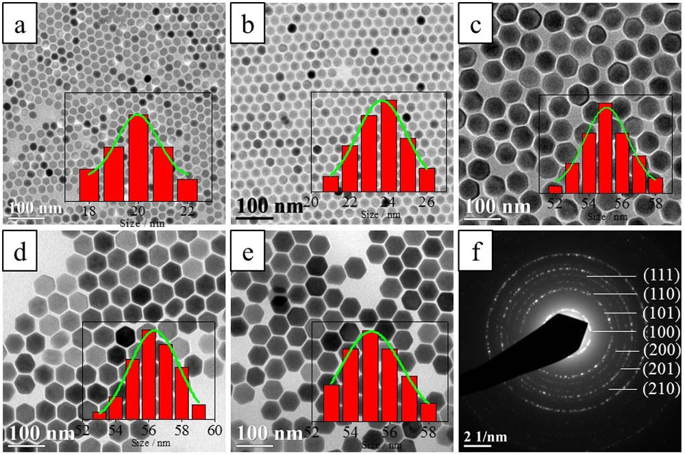
Two types of @ nanoparticles with incorporated ions were prepared to investigate the influence of the ions. In addition, 20%, 0.5% and 20%, 0.5%, 20% nanoparticles were also prepared as the contrastive samples. All samples were prepared by the procedure descripted in our previous work[34]. Figure 1 shows the typical transmission electron microscopy morphologies of such nanoparticles. The mono-dispersed 20%, 0.5%, 20% nanospheres exhibited an average size of , similar to that of the conventional 20%, 0.5% nanospheres, revealing that the incorporation had a minimal effect on the nanoparticle morphology and size. The @ nanoparticles exhibited a hexagonal shape with an average size of . The larger size of these nanoparticles was mainly due to the high concentration in the core. The two types of -incorporated @ nanoparticles were also hexagonal and showed a similar size to the @ nanoparticles. The selected-area electron diffraction (SAED) patterns of the @, nanoparticles confirmed their hexagonal shape with crystallographic phases belonging to the standard hexagonal host lattice (JCPDS 28-1192). All the results indicated that the incorporation in the @ nanoparticles did not affect the microstructure, morphology, or size, as in the case of the 20%, 1% nanoparticles.
Figure 1.Transmission electron microscopy morphologies of (a) 20%, 1%, (b) 20%, 0.5%, 20%, (c) @, (d) @@, (e) @, nanoparticles (the insets show the size distribution of the corresponding nanoparticles), and (f) SAED pattern of @ nanoparticles.
The photoluminescence spectra of the nanoparticles, with and without incorporated , were measured using a 980 nm laser excitation, as shown in Fig. 2. The photoluminescence intensity of 20%, 1%, 20% was reduced by 97%, although the ratio was enhanced from 0.9 to 7 compared with that of the 20%, 1% nanoparticles (Fig. 2(a)). This result revealed a strong quenching effect in the nanoparticles due to the incorporation. However, a slight effect of the incorporation on the emission intensity was observed in the @ nanoparticles, as shown in Fig. 2(b). Compared with the @ nanoparticles without incorporated , the photoluminescence intensity of the @ nanoparticles shelled by 20% was reduced by , and the ratio was approximately equal to 7, slightly higher than that of the @ nanoparticles. This result indicated that an enhancement of the red emission of the @ nanoparticles was not achievable by only coating them with a -contained shell layer. When the ions were incorporated into the shell of the @, nanoparticles, the ratio was enhanced to 11, nearly twice that of the @ nanoparticles, although the photoluminescence intensity was also reduced by . However, the emission intensity of the @, nanoparticles was enhanced by a factor of more than 120 compared with that of the 50%, 1%, 20% nanoparticles. These results demonstrated the weak quenching effect that exists in the @, nanoparticles.
Figure 2.Photoluminescence spectra of different types of ion incorporations: (a) 20%, 1% and (b) @ nanoparticles. (c) Photoluminescence intensity of the green and red emission bands. (d) The intensity ratios from the nanoparticles in (a) and (b).
The dependence of the intensities of the green and the red UC emission band on the pump power for the @, UCNPs was measured, as shown in Fig. 3. In general, the photoluminescence intensity increased on the pump laser power and obeyed the rule of , where is the photoluminescence intensity, is the pump laser power, and is the number of laser photons required. The slope values for the green and the red emission bands of the @, UCNPs approached 2, indicating that both the green and the red emissions involve a two-photon process for their generations. The slope values were in good agreement with previous results on -codoped UCNPs[32,34].
Figure 3.Logarithmic plot of the dependence of the intensities of the green and the red UC emission bands on the pump power for the @, UCNPs.
High emission intensity was observed in the core/shell nanoparticles consisting of a core containing ions and a shell containing and ions. This result implied that the strong quenching effect in the Yb/Ho/Ce-tridoped nanoparticles may be related to the interaction between the and ions. To verify and provide a theoretical background to this hypothesis, the steady-state rate equations were used. In this physical model, and are the population densities of the ions in the ground and the excited states, respectively; , , , , and are the population densities of the , , , , and states, respectively, of the ions; and and are the population densities of the ions in the ground and the excited states, respectively. In addition, , , and are the energy transfer rates from the excited ions to the ions, and , , , and are the radiation rates of the energy states of the ions. and are the phonon-assisted nonradiative relaxation rates from the to and from the to states, respectively, of the ions; and are the coefficients of the cross relaxations between and ions in the and states, respectively; and is the coefficient of the cross relaxation between the and ions. is the laser intensity at 980 nm, is the laser frequency, is the absorption cross section of the ion, is the radiation rate of the excited state of , and is the radiation rate of the excited state of . The steady-state rate equations for the discussed system can be described as follows: After solving Eqs. (1) and (2), we obtained the following expressions: Then, we can obtain the intensities of the red and green lights by applying the following equations: where and are the frequencies of the red light and green light, respectively. Furthermore, by combining Eqs. (5), (6), (9), and (10), we can derive the following expressions:
According to Eqs. (7) and (8), we know that
Moreover, considering Eqs. (11) and (12), we can also obtain the following expressions:
Hence, from Eqs. (14) and (15), it was demonstrated that both the intensities of the red and green lights decrease by the coefficient of the cross relaxation between and ions, although the two cross relaxations between and ions can lead to an enhancement of the red light radiation by reducing the green light radiation, as shown in Fig. 4(b). This model is able to explain the low emission intensity of the Yb/Ho/Ce-tridoped nanoparticles. When the and ions were moved to the shell layer, as shown in Fig. 4(c), the cross relaxation between the and ions could be reduced drastically as a result of the extended distance between the and ions, while the cross relaxation between the and ions was kept constant. Thus, high-intensity red light radiation was obtained for the @, nanoparticles. The reduction of the @, nanoparticles, compared with that of the @ nanoparticles (Fig. 2), mainly resulted from the cross relaxation between the and ions at the core/shell interface. The reduction of the @@ nanoparticles indicated that the quenching effect was further reduced by extending the distance between the and ions.
Figure 4.(a) Energy level diagrams of , , and ions and proposed UC mechanisms. (b) Schematic illustration of the proposed energy-transfer mechanisms in nanoparticles. (c) Schematic illustration of the proposed energy-transfer mechanisms in core/shell @ nanoparticles.
In conclusion, the ions are incorporated into the Yb/Ho-codoped nanoparticles to enhance the red emission. The incorporation enhances the intensity ratio between the red emission and the green emission of the nanoparticles, but largely reduces the total photoluminescence intensity. However, when the ions are incorporated into the shell of the core/shell @ nanoparticles, the emission intensity is also enhanced by a factor of more than 120 compared with that of the nanoparticles. This result indicates that the incorporation into the nanoparticles promotes a strong quenching effect that reduces the emission intensity; the quenching effect is significantly reduced by incorporating the ions into the core/shell structured Yb/Ho codoped nanoparticles. A theoretical model is proposed to explain the quenching effect existing in the nanoparticles, revealing that the quenching is mainly related to the interaction between the ions and the ions.