Jiangsu Key Laboratory of Advanced Laser Materials and Devices, School of Physics and Electronic Engineering, Jiangsu Normal University, Xuzhou 221116, China
The changes of mechanical properties and biological activities of monomeric erythrocytes are studied using optical tweezers micromanipulation technology. Firstly, the mechanical properties of irradiated erythrocyte membranes are obtained. Weaker power laser irradiation can delay the decay of the mechanical properties of erythrocytes and promote the biological activity of erythrocytes, while higher power laser irradiation damages erythrocytes. The stronger the laser irradiation is, the more obvious and rapid the damage will be. The temperature of the cell surface will be changed by regulating the laser power and irradiation time, so the biological functions of erythrocyte can be controlled. Secondly, the finite element simulation of the temperature change on the cell surface under the condition of laser irradiation is carried out using simulation software, and the precise temperature of the cell surface irradiated cumulatively by a laser with different powers is obtained. Finally, the processes of abscission, unfolding, and denaturation of hemoglobins in erythrocytes at different temperatures due to the photothermal effect are analyzed using the model. The mechanism of laser irradiation on the elasticity of erythrocyte membranes is also obtained.
With the development of laser technology, the laser medicine has been widely used in clinical applications. For example, a 755 nm alexandrite laser is used in the treatment of pigmentary lesions in Asians[1], Er-doped yttrium aluminum garnet (Er:YAG) and Er,Cr-doped yttrium scandium gallium garnet (Er,Cr:YSGG) lasers are used to ablate enamel and dentin[2], and intense pulsed light (IPL) is used in the treatment of evaporative-type dry eye disease[3]. In addition, laser medicine has many applications in the field of medical diagnosis. Photoacoustic imaging (PAI) is used to characterize the spatial and quantitative features of lipid-rich atherosclerotic plaques[4]. Optical coherence tomography (OCT) is used to investigate the cranial meninges in an animal model of brain injury in vivo[5]. Photoacoustic (PA) microscopy is used to achieve high-resolution, high-contrast, and large field of view imaging of skin[6]. In clinical applications, laser medicine is currently applied to the surface of organs and the blood in vessels. The use of laser radiation as an alternative energy-based method for sealing blood vessels is a typical clinical application[7]. Therefore, studying the photothermal biological effect of erythrocytes is of great significance for developing medical applications.
For blood microcirculation of the human body, the erythrocyte membrane should have good elasticity and plasticity when erythrocytes pass through the capillaries that have diameters smaller than erythrocytes[8]. The elasticity of erythrocytes is used to characterize the health of red blood cells in biophysics. Similarly, the elasticity of erythrocytes can be used to characterize the degree of laser irradiation on blood coagulation.
A large number of researchers have studied the effect of laser irradiation on the elasticity of erythrocytes. For example, Kujawa et al.[9] found that the 810 nm near-IR (NIR) laser could change the adenosine triphosphate (ATP) enzyme activity of the erythrocyte membrane ion pump, and the effect was related to the laser irradiation dose. Kovacs et al.[10] found that the membrane micro-viscosity of the erythrocytes reduced by half, and the fluidity of the membranes would significantly increase if erythrocytes were irradiated by a low-intensity He–Ne laser. Chowdhury et al.[11] found that the laser could rapidly damage the erythrocyte membranes when the laser power was larger than 280 mW, and the erythrocyte damage rate of patients with type II diabetes was different from normal erythrocytes. However, most of the researches have only studied the macroscopic physical and chemical properties of the erythrocyte population. It is necessary to deeply study the changes of mechanical properties of individual erythrocytes. It is quite difficult to reflect the changes of various functional parameters of a single erythrocyte in a complicated and changeable micro-environment. Optical tweezers have the advantages of single cell operation with higher precision and smaller biological damage[12–16].
Sign up for Chinese Optics Letters TOC. Get the latest issue of Chinese Optics Letters delivered right to you!Sign up now
For complex living organisms, accurate simulation calculations are essential. It can provide more comprehensive information for studying the mechanism of laser biological effects[17–19]. However, there are few reports about the effects of precise quantitative temperature on erythrocytes under laser irradiation. In addition, precise quantitative temperature at the cell level is difficult to measure by conventional methods. Therefore, it is necessary to accurately simulate the temperature change process of erythrocyte surfaces irradiated by lasers at different powers.
The experimental instrument was an acousto-optic deflector (AOD) scanning optical tweezers system (Tweez250si), including a Nikon Eclipse Ti inverted optical microscope and a 1064 nm Nd:YAG laser (the maximum output power is 5 W). The transmittance of the AOD is 36%, and the transmittance of the microscope objective is 70%. Laser power is regulated by the optical trap coefficient and laser level. The experimental samples were normal in vitro erythrocytes (conserved at 4°C) and phosphate buffered solution (0.01 mol/L). The erythrocyte solution was diluted with phosphate buffered saline (PBS), and the erythrocytes could non-specifically adhere to the bottom surface of the sample cell. Then, the sample was placed in the optical tweezer system, and the erythrocytes were stretched by an optical trapping force to observe the deformation of the cells and measure the parameters of mechanical properties of the cells. All experiments were performed at room temperature (20°C), and the experimental data were processed by TWV Viewer software[20].
The erythrocytes were stretched using the optical trap force, which induced erythrocyte deformation. As the optical trap moved outwards, the amount of deformation of the erythrocytes became greater, and then the erythrocytes would get rid of the optical trapping force and restore the original state when the stretching limit was reached. The maximum amount of deformation under a certain optical trapping force could be used to characterize the elasticity of erythrocytes and reflect the health of erythrocytes. The trap power was kept at 200 mW during the experiment to ensure the same tensile force. The radial length was recorded when the erythrocytes were stretched using the optical trapping force. Six groups of erythrocytes were selected, one of which was used as a non-irradiated control group, and each group data has average value. The powers of lasers irradiating erythrocytes were 0, 50, 100, 125, 150, and 200 mW. The erythrocytes were stretched every 1 min.
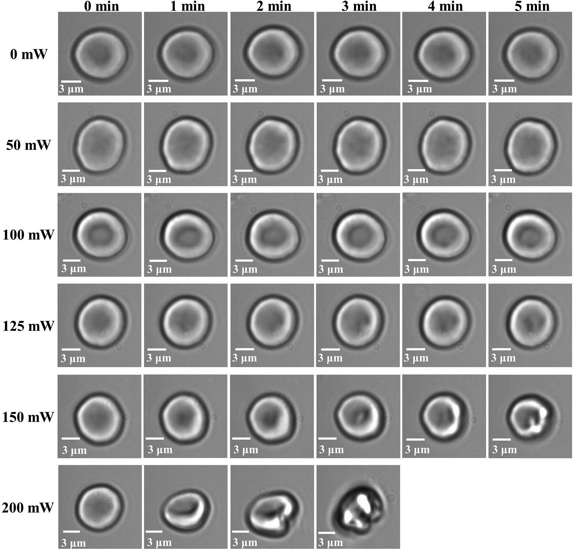
Figure 1 shows the states of erythrocytes irradiated by different laser power every 1 min. It can be seen that morphology of erythrocytes does not change much within 5 min without laser irradiation. The sizes of erythrocytes are slightly enlarged after 5 min when the laser power is 50 mW. When the laser power is larger than 50 mW, with the increasing of the laser irradiation time, the size of the erythrocytes becomes smaller, and it collapses at the center of irradiation, where the pixel becomes darker in the image. In the longitudinal direction, as the laser irradiation power increases, the erythrocytes decay faster. When the power is 150 mW, the erythrocytes hemolyze after 5 min. When the power is 200 mW, after 3 min, the erythrocytes begin to hemolyze.
Figure 1.States of erythrocytes under different power laser irradiation.
Figure 2 shows the state of erythrocytes under laser irradiation every 1 min. Without laser irradiation, there is almost no change for the amount of deformation of erythrocytes within 5 min. The amount of deformation of erythrocytes increases when laser power is 50 mW. When the laser power is larger than 50 mW, with the increasing of the laser irradiation time, the amount of deformation of erythrocytes becomes smaller. With the increasing of the laser power, the amount of deformation of erythrocytes decreases faster and faster. The erythrocytes begin to hemolyze when the laser irradiation time is 5 min and the laser irradiation power is 150 mW. While the erythrocytes begin to hemolyze when the laser irradiation time is 3 min, and the laser irradiation power is 200 mW, then the three-dimensional shapes of erythrocytes are completely defocused, and the erythrocytes are unable to be stretched.
Figure 2.Elongation of erythrocytes under different power laser irradiation.
There are individual differences among the erythrocytes used in the experiment. So, it is necessary to normalize the data and calculate the relative elongation of the erythrocytes: where is the initial size of erythrocytes, is the maximum stretched length, is the elongation, is the elongation in the initial state, and is the relative elongation. Under different irradiation power, the relative elongations of the erythrocytes irradiated by lasers with different irradiation times are calculated, which are shown in Fig. 3.
Figure 3.Relationship between the relative elongation of erythrocytes and irradiation time under different power.
In Fig. 3, when the laser power is 50 mW, the relative elongation . In other words, the elasticity of erythrocytes is superior to erythrocytes that have not been irradiated with a laser. When the erythrocytes are irradiated by a low-power laser, the relative elongation becomes larger than that of the non-irradiated state, and the elasticity increases, which indicates that the low-power laser has a certain positive promoting effect on the erythrocytes. When the erythrocytes are irradiated by a laser whose power is higher than 50 mW, the relative elongation of erythrocytes decreases with increasing power and irradiation time. Then, the erythrocyte membranes rupture, and eventually erythrocytes hemolyze. This shows that high-power lasers have destructive effects on erythrocytes, and, with the increasing of laser power, the time required to destroy erythrocytes is shorter and shorter.
Since the surface temperature of erythrocytes cannot currently be measured directly, the temperature changes of the surface of a monomeric erythrocyte under laser irradiation are simulated. The main ingredients of erythrocytes are the cell membranes and cytoplasm, so the erythrocytes can be simplified into empty shells[21]. Using Comsol Multiphysics software, the cytoplasm is simplified to water because it is similar to water. The cell membrane thermal conductivity is set to , the density is set to , and the constant pressure heat capacity is set to . Different power lasers are used during the simulation, and the simulation time is set to 5 min, which is consistent with the experimental time.
As shown in Fig. 4, when the optical power is set to 150 mW, the temperature of the central irradiation point of the erythrocytes is constantly rising, and the temperature isosurface is continuously expanding. After 5 min, the temperature of the erythrocytes rises to 67°C.
Figure 4.Temperature changes of a monomer erythrocyte under 150 mW laser irradiation.
The same simulation method is used to simulate the temperature rising of erythrocytes under different laser irradiation. The simulation results are shown in Fig. 5.
Figure 5.Surface temperatures of erythrocytes under different power lasers changing with time.
From the Fig. 5, as the irradiation time increases, the surface temperature of the erythrocytes rises. With the increasing of the laser power, the temperature rises faster. When the laser irradiation power is at 50 mW, the temperature of a monomer erythrocyte reaches 37°C after 5 min, and 37°C is the best temperature for the activity of erythrocytes[22,23]. When the laser has a high power of 200 mW, the erythrocyte reaches a temperature of 83°C after 5 min, which exceeds the tolerance temperature of erythrocytes[24–27]. Combining with the relative elongation of erythrocytes under different irradiation times, the relationship between the cell surface temperature and the relative elongation under different laser powers is shown in Fig. 6.
Figure 6.Changes in surface temperature and relative elongation of erythrocytes under different power lasers.
Under weak power laser irradiation, the temperature of erythrocytes rises from room temperature (20°C) to body temperature (37°C), the activity of erythrocytes increases, and their elasticity becomes stronger. The weak power laser produces a positive promotion for the erythrocytes. However, under strong laser irradiation, rising temperatures denature hemoglobins, which leads to a decreasing of the elasticity of membranes. When the temperature reaches 50°C, hemoglobins begin to unfold. The denatured hemoglobins are insufficient to support the erythrocyte membrane structure, which causes the erythrocyte membranes to collapse or even rupture and hemolyze. Moreover, it can be seen from Fig. 6. that with the increasing of temperature, the elasticity of the erythrocyte membranes decreases quickly, and the erythrocytes are destroyed more rapidly.
Laser irradiation of biological cells can produce five biological effects[28–30], namely, the thermal effect, photochemical effect, mechanical effect, electromagnetic field effect, and biostimulation. The optical wavelength used in this experiment is 1064 nm, which belongs to the NIR range, and its biological heating effects are stronger. Therefore, when the laser irradiates erythrocytes, the mechanical properties of the erythrocytes change because of the temperature change caused by the thermal action.
A cytoskeleton (composed by different proteins) plays the most important role in the shape and elasticity of cells. Low-intensity laser irradiation activates superoxide dismutase (SOD), increases SOD activity, and effectively removes free radicals from the surface of erythrocyte membranes[31]. Moreover, low-intensity laser irradiation can activate the erythrocyte membrane -ATP enzyme[32], release ATP energy to erythrocytes, and ensure that erythrocytes have enough energy to maintain cell morphology and deformability. The ability of erythrocytes to deform is thus improved. As the laser irradiation power increases and the irradiation time increases, the accumulation of heat causes an increase in temperature and affects the erythrocytes[28]. Photothermal action affects the enzymes on the erythrocyte membrane. When the laser irradiation temperature reaches a certain value, the enzyme denatures and affects the activity of erythrocytes. High temperatures denature the membrane proteins of erythrocytes, causing damage to the membrane skeleton proteins. Strong short-term laser-induced local overheating can cause cell damage[33]. The deformability of erythrocytes is thus reduced.
Laser irradiation affects the erythrocyte membranes; after this, the denaturation of hemoglobin occurs in a second stage. The thermal denaturation process of hemoglobin consists of three distinct phases[34]. During 30–44°C, the hemoglobins are in the structural perturbation stage, corresponding to 50 mW weak power laser irradiation in our experiment. The deformability of erythrocytes is related to the amount of hemoglobins adhering to the erythrocyte membranes. The larger the amount of hemoglobins is, the worse the deformability of erythrocytes is[35]. Hemoglobins can play a role in supporting the cytoskeleton. Because the connection between the hemoglobin and the membrane is a weakly coupled interaction[36], during this phase, hemoglobins absorb energy and then fall off into free hemoglobins, which leads to a certain increase of the erythrocytes deformability. During 44–54°C, the hemoglobins are in the thermal unfolding stage, corresponding to the laser irradiation of 100 mW in our experiment. At this stage, the secondary bonds (hydrogen bonds and salt bonds), the connecting peptide chain in hemoglobins, are broken under photothermal action, causing the protein to unfold, then the erythrocyte membrane becomes brittle, and the elasticity of the erythrocyte membrane decreases. During 54–70°C, the hemoglobins are in the thermal aggregating stage, corresponding to the high-power laser irradiation of 200 mW in our experiment. At this stage, the secondary bonds are further broken, hemoglobins are denatured, and the erythrocyte membranes are broken. The structural change processes of the erythrocyte membrane and hemoglobin bilayer model are shown in Fig. 7.
Figure 7.(a) Initial state of the erythrocyte membrane and hemoglobin bilayer model. (b) Process of denaturation of the hemoglobin layer. (c) Final state of the hemoglobin layer.
According to Hooke’s law, a two-layer structure model of the erythrocyte membrane and hemoglobin is established: where is the elastic coefficient of the two-layer structure model of the erythrocyte membrane and hemoglobin, is the elastic coefficient of the hemoglobin layer, is the elastic coefficient of the membrane layer, and is the number of hemoglobins. When the hemoglobins are denatured, the amount of hemoglobins in the hemoglobin layer decreases, and the elastic coefficient of the two-layer structure increases; so, the brittleness of the membrane increases, and the elasticity decreases. Eventually, the cell membranes rupture, and the cells hemolyze.
In this Letter, optical tweezers micromanipulation technology is used to stretch monomeric erythrocytes, and the mechanical properties and biochemical activities of erythrocytes under different laser irradiation are studied. By adjusting the laser power and irradiation time, the mechanical properties of erythrocyte membranes under different cumulative laser irradiation are studied. Weaker power laser irradiation promotes the biological activity of erythrocytes, while higher power laser irradiation damages erythrocytes. The temperature changes of the surface of erythrocytes irradiated by laser affects the mechanical properties and biological activity of erythrocytes. Through the simulation using the software, under laser irradiation, the precise change of the surface temperature of a monomeric erythrocyte is obtained. Weaker power laser irradiation causes a slow temperature rise, while a higher power laser causes a fast temperature rise. A double-layer model of the erythrocyte membrane and hemoglobin is established. Based on the photothermal effect, the processes of shedding, unfolding, and denaturation of hemoglobins in erythrocytes at different temperatures are analyzed. The mechanism of the laser irradiation on the elasticity of erythrocyte membranes is also obtained. Denatured hemoglobins reduce the elasticity of the erythrocyte membrane. The results of the study can be used to judge the activity of erythrocytes and characterize the functional status index. It provides a valuable reference for clinical health care and even cell therapy.