• Chinese Optics Letters
  • Vol. 21, Issue 10, 103601 (2023)
Tianyue Zhang1、2、*, Zhiyuan Wang1, Xiangchao Zhong1, Ying Che1, and Xiangping Li1、**
Author Affiliations
  • 1Guangdong Provincial Key Laboratory of Optical Fiber Sensing and Communications, Institute of Photonics Technology, Jinan University, Guangzhou 510632, China
  • 2School of Integrated Circuits, Beijing University of Posts and Telecommunications, Beijing 100876, China
  • show less
    DOI: 10.3788/COL202321.103601 Cite this Article Set citation alerts
    Tianyue Zhang, Zhiyuan Wang, Xiangchao Zhong, Ying Che, Xiangping Li. Photothermal nonlinear scattering of shell-isolated gold nanoparticles and applications in super-resolution imaging[J]. Chinese Optics Letters, 2023, 21(10): 103601 Copy Citation Text show less
    (a) Schematic illustration of the different local thermal environments created for 70-nm-diameter gold nanospheres. (b) Experimental measurements of nonlinear plasmonic scattering of the five prepared samples, showing the nonlinear dependency of scattering on irradiance intensities for continuous-wave excitations at the wavelength of 532 nm. The optical setup of the reflected laser scanning confocal microscope is shown in the inset. BS, beam splitter; OL, objective lens; PMT, photomultiplier tube.
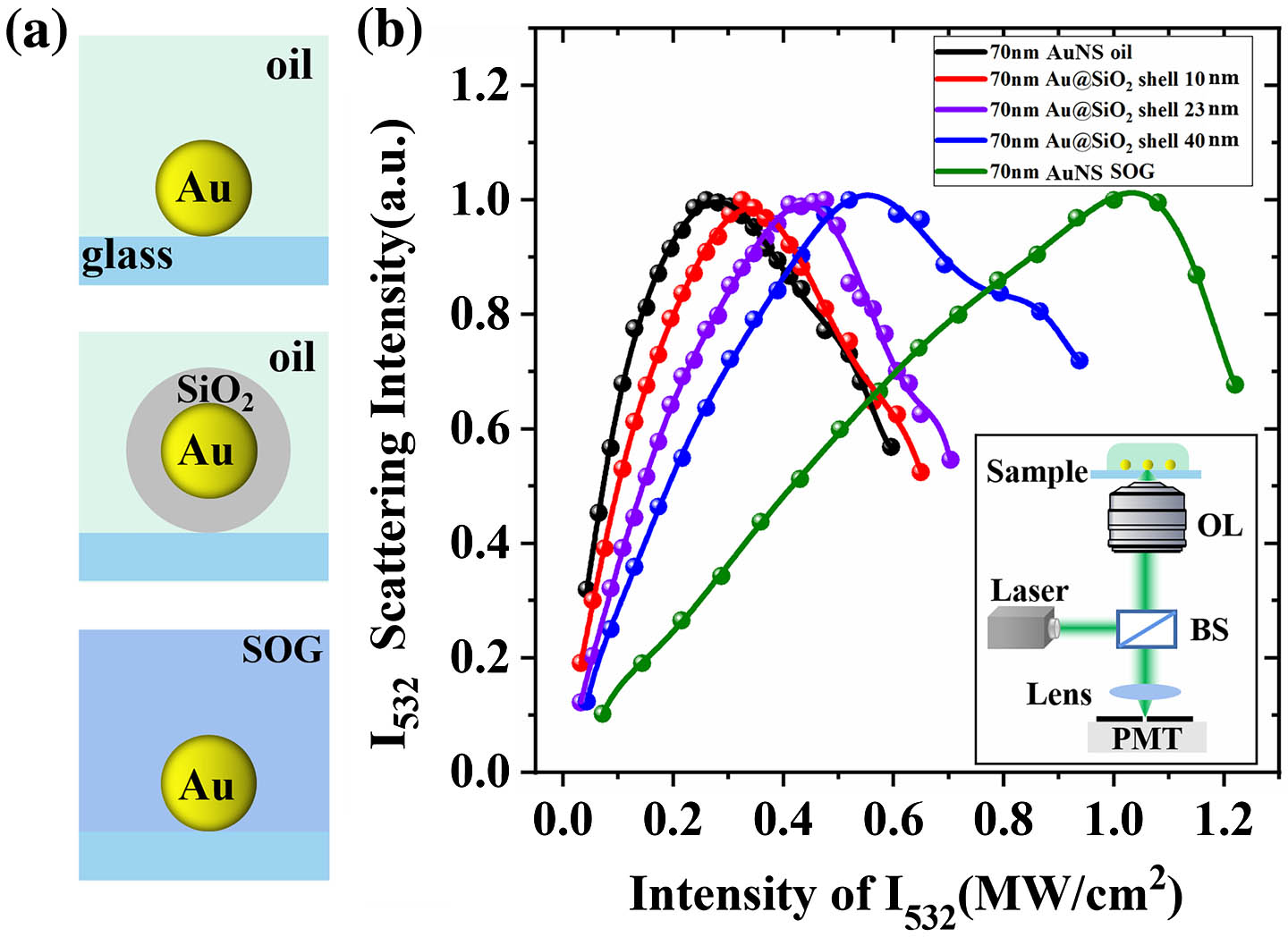
    Fig. 1. (a) Schematic illustration of the different local thermal environments created for 70-nm-diameter gold nanospheres. (b) Experimental measurements of nonlinear plasmonic scattering of the five prepared samples, showing the nonlinear dependency of scattering on irradiance intensities for continuous-wave excitations at the wavelength of 532 nm. The optical setup of the reflected laser scanning confocal microscope is shown in the inset. BS, beam splitter; OL, objective lens; PMT, photomultiplier tube.
    (a) Illustration of optical heating of the gold nanospheres that converts light into temperature rises. Due to the difference in the local thermal media, the gold nanospheres (top, in immersion oil; middle, in SOG materials; and bottom, with silica coatings) undergo various temperature rising. (b) Theoretical calculations of the temperature evolution of a single 70-nm-diameter gold nanosphere surrounded by different thermal host media characterized by κeff and subject to CW laser illumination with increasing laser intensity I532. (c) The evolution of the scattering cross sections of the gold nanospheres with excitation intensities. (d) The calculated plasmonic scattering intensities of the gold nanospheres as a function of 532 nm laser intensities I532 at a different κeff.
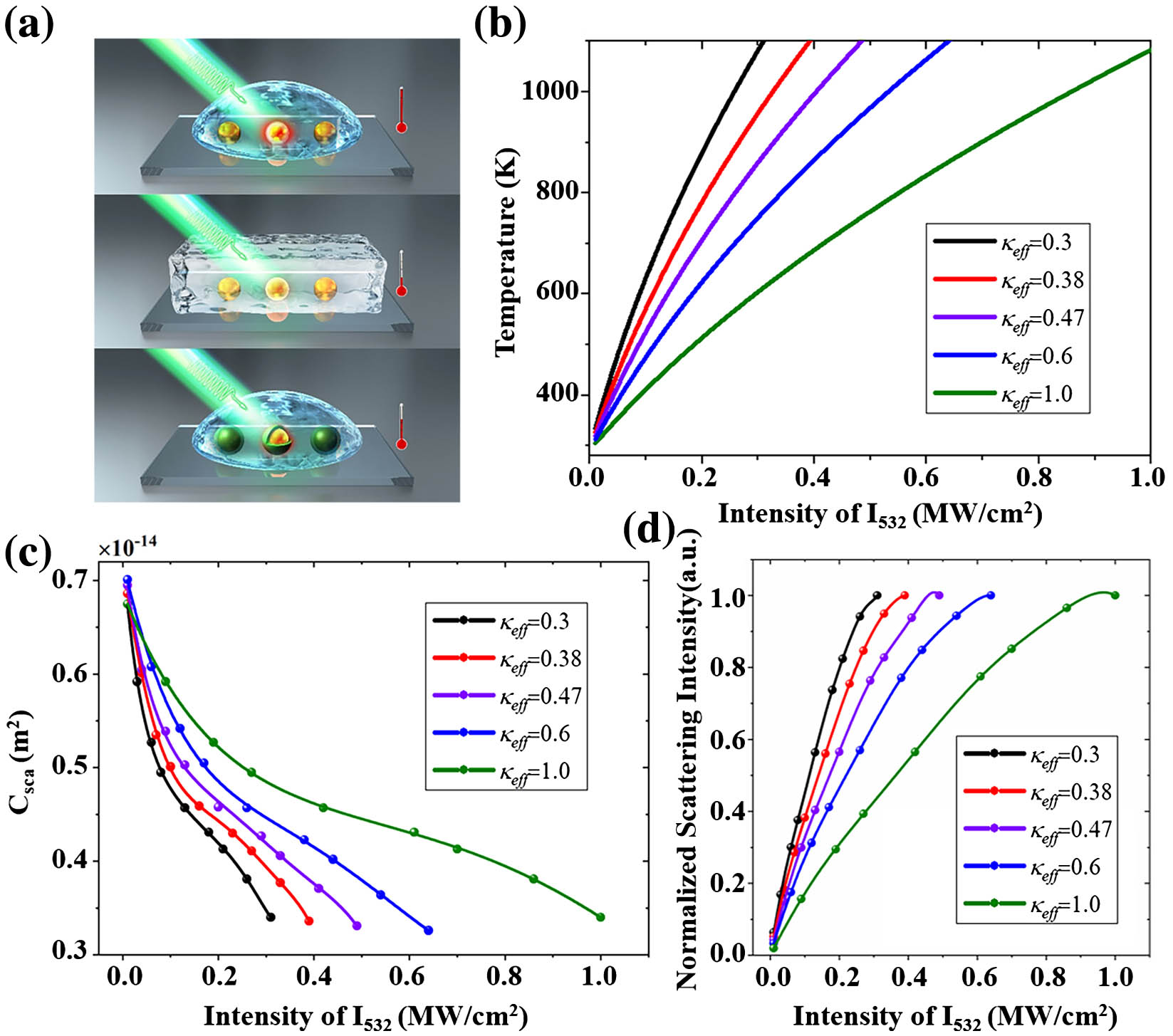
    Fig. 2. (a) Illustration of optical heating of the gold nanospheres that converts light into temperature rises. Due to the difference in the local thermal media, the gold nanospheres (top, in immersion oil; middle, in SOG materials; and bottom, with silica coatings) undergo various temperature rising. (b) Theoretical calculations of the temperature evolution of a single 70-nm-diameter gold nanosphere surrounded by different thermal host media characterized by κeff and subject to CW laser illumination with increasing laser intensity I532. (c) The evolution of the scattering cross sections of the gold nanospheres with excitation intensities. (d) The calculated plasmonic scattering intensities of the gold nanospheres as a function of 532 nm laser intensities I532 at a different κeff.
    (a) The FWHM of the scattering image spots using bare AuNSs and 40-nm-shell-coated AuNSs, with increasing suppression beam intensity I532. A series of images with various suppression beam powers were taken, allowing for the quantification of the resolution dependence on the applied suppression power. The mean spot sizes for each power setting were determined by measuring the FWHM of the Gaussian-fitted profiles of individual nanoparticles and averaging over ten particles. The SEM of bare gold and Au@SiO2 dispersed on the ITO substrate was performed for characterization. The SEM images were also shown in the figure. The region enclosed by the shaded area only involves the SUSI of 40-nm-shell-coated AuNSs. For the intensity larger than 0.8 MW/cm2, the bare AuNSs were no longer thermally stable. (b) Confocal and SUSI microscopy imaging of isolated 40-nm-shell-coated AuNSs. (c) The intensity lateral profile of the selected nanoparticles (numbered by I and II). Discrete circle points are measured intensity profiles, and the solid lines are Gaussian fits.
    Fig. 3. (a) The FWHM of the scattering image spots using bare AuNSs and 40-nm-shell-coated AuNSs, with increasing suppression beam intensity I532. A series of images with various suppression beam powers were taken, allowing for the quantification of the resolution dependence on the applied suppression power. The mean spot sizes for each power setting were determined by measuring the FWHM of the Gaussian-fitted profiles of individual nanoparticles and averaging over ten particles. The SEM of bare gold and Au@SiO2 dispersed on the ITO substrate was performed for characterization. The SEM images were also shown in the figure. The region enclosed by the shaded area only involves the SUSI of 40-nm-shell-coated AuNSs. For the intensity larger than 0.8 MW/cm2, the bare AuNSs were no longer thermally stable. (b) Confocal and SUSI microscopy imaging of isolated 40-nm-shell-coated AuNSs. (c) The intensity lateral profile of the selected nanoparticles (numbered by I and II). Discrete circle points are measured intensity profiles, and the solid lines are Gaussian fits.
    Correlated super-resolution SUSI and SEM images for (a), (b) two closely spaced bare AuNSs and (c)–(e) pairs of 40-nm-shell-coated AuNSs. The gap-to-gap distances of two nanoparticles are shown in the SEM images. The super-resolution imaging of the shell-isolated nanoparticles clearly shows the resolving ability to distinguish two nanoparticles that are closely located in sub-diffraction spaces as small as 5 nm. The rightmost column plots the cross section profiles of the scattering images in confocal and SUSI.
    Fig. 4. Correlated super-resolution SUSI and SEM images for (a), (b) two closely spaced bare AuNSs and (c)–(e) pairs of 40-nm-shell-coated AuNSs. The gap-to-gap distances of two nanoparticles are shown in the SEM images. The super-resolution imaging of the shell-isolated nanoparticles clearly shows the resolving ability to distinguish two nanoparticles that are closely located in sub-diffraction spaces as small as 5 nm. The rightmost column plots the cross section profiles of the scattering images in confocal and SUSI.
    Tianyue Zhang, Zhiyuan Wang, Xiangchao Zhong, Ying Che, Xiangping Li. Photothermal nonlinear scattering of shell-isolated gold nanoparticles and applications in super-resolution imaging[J]. Chinese Optics Letters, 2023, 21(10): 103601
    Download Citation