Optical micro-endoscopes are widely used in various biomedical and clinical applications. As a semi-invasive imaging tool, it can detect and image the tissues and viscera of the human body (or other animals), and achieve the visualization of hard-to-reach interior areas. It can also be used to study the brain activities of free-moving animals. In order to reduce the invasive damage, it is usually necessary to limit the size of the endoscopic probe, which will lead to the degradation of imaging performance, especially the axial focusing ability and three-dimensional imaging ability, limiting its important role in disease diagnosis and in vivo observation.
Existing optical micro-endoscopes cannot achieve high imaging quality, large detection depth, and small probe size simultaneously, which makes it difficult for them to go deep into the small and narrow parts of the human body, such as the micro-vessels, alveoli, and cochlea, as well as the intestines and stomachs of small animals. In addition, in the conventional gradient index lens (GRIN) endoscope system and optical fiber endoscope system, micro electro mechanical systems (MEMS) or micro optoelectronic components are usually required to obtain three-dimensional information, whether through axial scanning or refocusing control, increasing the cost and complexity of the system.
In recent years, the single-mode fiber bundle has been used to develop compact and convenient endoscopic imaging systems, avoiding reliance on bulky mechanical or optoelectronic components. With a detection length of tens of centimeters, tens of thousands of single-mode fiber cores arranged in a diameter of several micrometers, and bendable materials, it provides a new idea for achieving high-performance three-dimensional (3D) endoscopic imaging. However, the existing methods have not fully utilized the large spatial bandwidth product (SBP) of fiber bundles, and it is still a challenge to achieve high-performance epi-fluorescence 3D imaging in a compact system.
To address this problem, the research group led by Prof. Xun Cao at Nanjing University in collaboration with Prof. Qionghai Dai at Tsinghua University has jointly constructed an all-optical endoscope probe by using a single-mode fiber bundle and a small-size light field imaging mode, realizing a snapshot 3D epi-fluorescence endoscopic imaging system with high imaging quality, large detection depth (~50 cm) and micro probe size (2 mm diameter). Relevant research results were published in Photonics Research, Volume 10, No. 9, 2022 (You Zhou, Bo Xiong, Weizhi Song, Xu Zhang, Guoan Zheng, Qionghai Dai, Xun Cao. Light-field micro-endoscopy using a fiber bundle: a snapshot 3D epi-fluorescence endoscope[J]. Photonics Research, 2022, 10(9): 2247).
The method reported in this article realizes 3D epi-fluorescence endoscopic imaging at camera frame rate by using the combination of a single-mode fiber bundle, a small micro-lens array (MLA), and a small lens pair, and further improves the imaging performance by conducting optical system optimization and applying dictionary learning based algorithm. Flexible and bendable fiber bundles also provide more convenience and flexibility in practical operation.
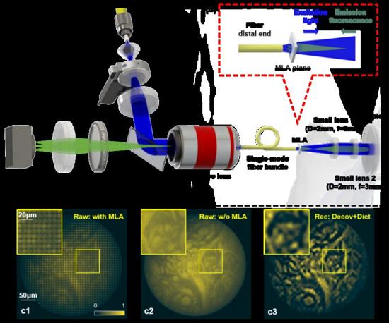
Fig. 1. (a) schematic diagram of the snapshot 3D endoscopic imaging system; (b) epi-fluorescence imaging model, and the micro-lens array is placed at the end of the optical fiber to obtain depth information; (c) imaging performance of human skin tissue.
As shown in Fig. 1, a single-mode fiber bundle is used to relay the light from hard-to-reach areas of the endoscopic scene. A 2-mm diameter MLA is placed at the distal fiber tip to obtain the depth information. The small imaging lens pair is applied to provide 3-fold magnification imaging.
Figure 2 shows the 3D imaging performance of the proposed endoscopic imaging method on biological samples. We compare different reconstruction results using the traditional Richardson–Lucy deconvolution method and the dictionary learning based method. The experimental results show that the dictionary learning based method can significantly reduce the reconstruction artifacts, enhance the imaging contrast, and improve the axial imaging accuracy. At the bottom of Fig. 2, we draw the line profile of two spots in HeLa cell samples, having different axial positions. We can detect their axial distance is about 25.95 microns, demonstrating the 3D imaging ability of the proposed method. By imaging the randomly distributed fluorescent beads, human skin tissues, HeLa cells, and other scenes, our method demonstrates an achievable imaging performance of 333-micron field of view, more than 24-micron depth of field, and up to about 4-micron lateral resolution.
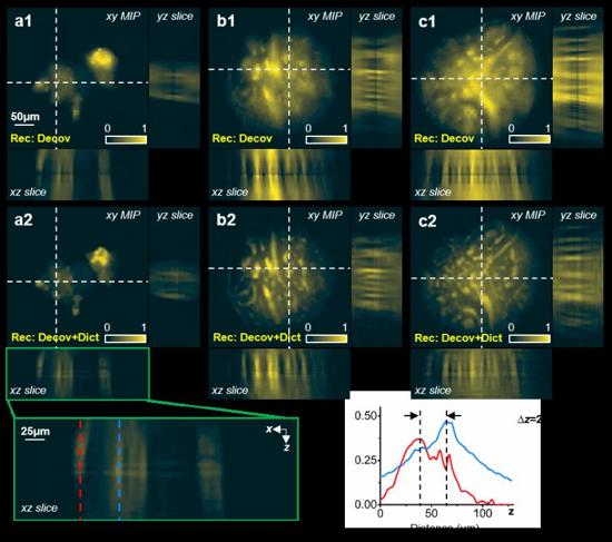
Fig. 2. 3D imaging performance by the proposed endoscopic system. (a1, a2) HeLa cell imaging recovered using Richardson–Lucy deconvolution (decov) and dictionary learning (decov + dict) respectively; (b1 ~ c2) human skin tissue imaging using these two methods.
"Combined with further system integration optimization and intelligent reconstruction algorithm, the cooperative team is expected to break through the limitations of the current system and realize the real-time 3D fluorescence visualization of the hard-to-reach interior in small and narrow tissues and organs of the human body or model animals in vivo", said by Prof. Xun Cao.