
Chao Liu, Jingwei Lü, Wei Liu, Famei Wang, Paul K. Chu. Overview of refractive index sensors comprising photonic crystal fibers based on the surface plasmon resonance effect [Invited][J]. Chinese Optics Letters, 2021, 19(10): 102202
- Chinese Optics Letters
- Vol. 19, Issue 10, 102202 (2021)
Abstract
1. Introduction
Surface plasmon resonance (SPR) is a critical physical–optical phenomenon involving excitation of electron density oscillations at metal-dielectric interfaces under the irradiation of -polarized light waves[
Driven by the large demand for miniaturization and integration and because optical fibers can transmit light waves based on total internal reflection, optical fibers constitute a desirable platform to excite SPR when plasmonic materials are coated on the surface of the fiber core[
The recent development of optical fiber sensors coincides with the rapid advance of micro/nano-fabrication technology giving rise to novel photonic crystal fibers (PCFs)[
Sign up for Chinese Optics Letters TOC. Get the latest issue of Chinese Optics Letters delivered right to you!Sign up now
Owing to the large impact of the technology, many papers have been published on PCF-SPR sensors, and our current knowledge is that the proper design and optimization determine the sensing properties of PCF-SPR sensors, for example, in bio-sensing and temperature and concentration measurements. In addition to Zhao et al., who summarized the progress of PCF-SPR sensors in 2017 and completed an overview of chemical sensors[
2. Physical Principle of SPR
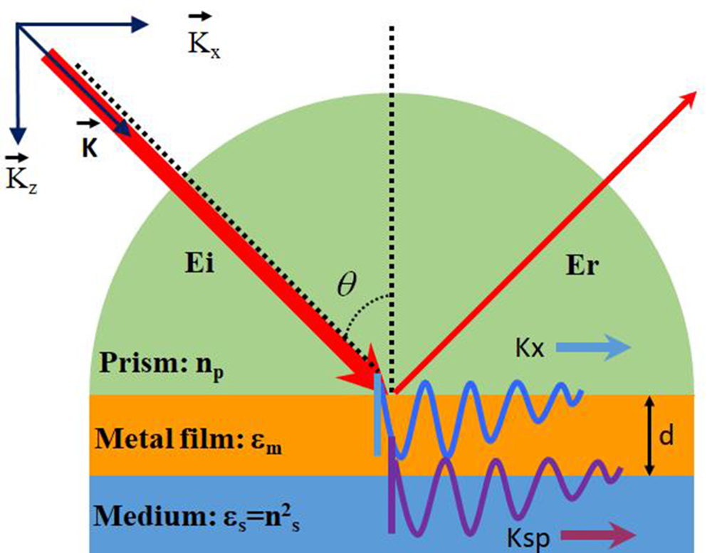
SPR is a phenomenon about optical excitation of surface plasmon waves (SPWs) by the method of attenuated total reflection except electron excitation. Before discussing PCF-SPR sensors, the SPR theory is described. The prism-structure model proposed by Otto[
![]()
Figure 1.SPR excitation by prism coupling using the Kretschmann configuration.
2.1. Surface plasmon wave
The SPW is a charge-density oscillation that exists at the interface between a metal thin film and a dielectric substrate with dielectric constants of opposite signs. The wave vector of SPW reaches the maximum value at the interface and decays evanescently into both media. The magnetic vector of the SPW is perpendicular to the propagation direction of the SPW and parallel to the plane of interface, the so-called TM-polarized wave. The propagation constant () of SPW propagating at the interface between the dielectric and metal film can be defined as[
2.2. SPR excitation by the evanescent wave
The evanescent wave is central to SPR sensing, and it is conveniently considered based on the total internal reflection. Supposing an electromagnetic plane wave propagates in a surrounding medium, which is expressed mathematically by electric field ,
The amplitude of the electric field () decays exponentially along the vertical direction. The electric field propagates in the direction along the metal surface, and it is called the evanescent wave. The propagation constant of the evanescent wave is given by the following expression:
SPW is not excited by an arbitrary light beam, and the phase matching conditions must be satisfied. It is a TM-polarized wave, so only TM polarized waves may excite SPW. The dispersion relation of SPW exhibits a nonlinear characteristic, and the momentum of the SPW is larger than that of light in free space for the same frequency, resulting in momentum mismatch between the light and SPW. The mismatch may be overcome by the evanescent wave from the prism coupler. In the prism configuration, the dispersion relation for the light wave and metal shows an intersection and therefore, SPW may be excited by the coupling light wave, as shown in Fig. 2.
![]()
Figure 2.Dispersion relation of TM incident light coupling with SPP.
In the three-layer system 0/1/2: prism/metal/medium geometry, as shown in Fig. 1, the reflected intensity is determined by Fresnel equations and the reflectivity for -polarized light, with the incident and the reflected field, is given by Eq. (6)[
When the phase matching point between the plasmon wave and evanescent wave is satisfied according to Eq. (9), a dip occurs in the reflectivity curve, as shown in Fig. 3. The relationship between the resonance wavelength/angle and RI of the analyte can be described, for example, as shown in Fig. 3(b), to enable SPR sensing of the analyte. From the perspective of practical applications, a linear relationship between them is preferred:
![]()
Figure 3.Methodology of SPR measurement: (a) SPR spectra for different RIs and (b) relationship between the wavelength and RI.
3. Simulation, Characteristics, and Fabrication of PCF-SPR Sensors
3.1. Simulation method for PCF-SPR sensors
Theoretical simulation is an effective and powerful tool to evaluate the sensing performance of sensors before actual fabrication. Since PCF-SPR sensors are complicated owing to the large number of design parameters and degree of energy coupling between the fundamental core mode and plasmonic mode, it is crucial to understand the detailed influence of the structural parameters of the PCF and plasmonic materials on the optical properties of the sensors in order to accurately predict the sensing characteristics. The design of a typical PCF-SPR sensor involves at least a dozen parameters such as the air hole size, plasmonic-layer thickness, and fiber core diameter. PCF-SPR simulation includes the process of selecting the optimal design and using models to predict the sensing performance for each structural combination. The primary advantage of combining computer simulation and modal analysis is that the modeler can try different approaches to obtain the solution for the same problem in order to design and optimize structural parameters of the desired PCF-SPR sensors.
According to the physics of SPR, when the phase matching conditions in PCF-SPR sensors are achieved, the largest energy is transferred from the fundamental core-guided mode to the plasmonic mode, leading to loss of the transmitted energy in fibers. Mathematically, the confinement loss is often employed to describe the sensing properties of PCF-SPR sensors, which is expressed as[
In order to assess the sensing performance of PCF-SPR sensors, there are three important parameters: spectral sensitivity, amplitude sensitivity, and RI resolution. The spectral sensitivity [] is defined as[
The amplitude sensitivity () is expressed as[
Many plasmonic PCF-SPR structures have been simulated by various methods, and the finite element method (FEM) is the most common method to compute the optical properties of PCF-SPR sensors. By means of FEM, Hassani et.al.[
![]()
Figure 4.(a) Loss spectra of two PCFs with and without defect. (b) Electronic field distributions of the PCF with defect: insets (c) and (d) show the electronic field distributions of the fundamental core-guided mode and higher-order plasmonic mode at λ = 575 nm; insets (e) and (f) show the energy distribution of the fundamental core-guided mode and fundamental plasmonic mode at λ = 632 nm[
Although SPR simulation can be carried out by means of Rsoft, Opti-FDTD software based on the finite-difference time-domain (FDTD) method, FDTD requires solving the partial differential equations in the time domain, consequently requiring a fine mesh and occupying a great deal of computer memory[
3.2. Performance of PCF-SPR sensors
PCF-SPR sensors have attracted considerable interests, and various sensing structures have been demonstrated to deliver excellent sensing performance. The properties are described based on the following categories: (1) RI detection including low/high RIs, (2) controllable resonance wavelength, and (3) extremely high sensitivity.
3.2.1. Low refractive index detection
The distinguishing feature of SPR is extremely high sensitivity to variations in RI of the surrounding medium, and sensing applications of SPR originate from the changes of the RI caused by the measured parameters. Hence, RI monitoring is one of the most significant functions of PCF-SPR sensors and also fundamental to the measurement of other parameters such as temperature, concentration, and magnetic field.
One of the crucial issues for SPR effect excitation is to satisfy phase matching conditions between the fundamental core-guided mode and plasmonic mode. Mathematically, phase matching is fulfilled when the effective RIs of the above-mentioned two modes are equal at a given wavelength. Generally, the effective RI of the fundamental core-guided mode in a PCF is close to that of the silica core, which is higher than 1.45 in practical applications. According to the Eq. (1), the real part of the effective RI of the SPW is approximately close to that of the surrounding medium[
Generally, the upper limit of RI detection for a PCF-SPR sensor is less than or equal to 1.33, and it is defined as a low RI sensor. At present, there are many applications available that need to detect a lower RI, such as aerogel[
![]()
Figure 5.Electronic field distributions of the sensor for different modes: (a) for the core-guided mode, (b) for the plasmonic mode, and (c) for the two modes at resonance point[
Haque et al.[
![]()
Figure 6.(a) Schematic illustration of the side-polished sensor. (b) Dispersion relations and loss spectra of the sensor. (c) Resonant curves for RIs of 1.00–1.20. (d) Resonant curves for RIs of 1.21–1.37[
3.2.2. High refractive index detection
The lower limit of RI detection greater than 1.33 is referred to high RI PCF-SPR sensing. High RI detection is useful in determining chemo-analytes and bio-analytes such as polymethylphenyl siloxane, glucose, benzene, and DNA[
![]()
Figure 7.(a) 2D diagram of the sensor. (b) Three-dimensional (3D) diagram of the sensor. (c) Dispersion relations and loss spectrum of the sensor (red line represents channel 1 for na = 1.35, green line represents channel 2 for na = 1.38). (d) Electric field distributions of the sensor for different wavelengths[
Besides graphene, semiconductor oxide- has also obtained great attention due to the enhancement for the SPR effect[
| Ref. | Characteristic | Refractive Index Range | Max. Sensitivity (nm/RIU) | Resolution (RIU) | Str. Diagram |
|---|---|---|---|---|---|
| [88] | Birefringence PCF | 1.00–1.43 | 6300 |  | |
| [76] | D-shaped PCF | 1.18–1.36 | 20,000 |  | |
| [90] | Concave-shaped PCF | 1.19–1.29 | 10,700 |  | |
| [60] | D-shaped PCF | 1.20–1.29 | 11,055 |  | |
| [66] | Microchannel PCF | 1.22–1.37 | 51,000 |  | |
| [62] | Gold-nanowire-coated PCF | 1.27–1.36 | 6000 |  | |
| [77] | D-shaped PCF | 1.27–1.32 | 10,493 |  | |
| [91] | Gold-coated PCF | 1.29–1.49 | 4156 |  | |
| [92] | PCF with circular air holes | 1.32–1.43 | 30,500 (x), 41,500 (y) | (x), (y) |  |
| [40] | PCF with exposed core | 1.33–1.42 | 13,500 |  | |
| [78] | Graphene-based D-shaped PCF | 1.33–1.37 | 3700 |  | |
| [93] | Copper-graphene-based PCF | 1.33–1.37 | 2000 |  | |
| [94] | External gold-layer-coated PCF | 1.33–1.37 | 4000 |  | |
| [95] | Hexagonal sensor | 1.33–1.42 | 11,000 |  | |
| [96] | PCF with exposed core | 1.33–1.42 | 16,400 |  | |
| [97] | Au-coated dual-core PCF | 1.33–1.51 | 6021 |  | |
| [98] | Hollow core Ag-coated PCF | 1.36–1.37 | 4200 |  | |
| [79] | D-shaped PCF coated with gratings | 1.36–1.38 | 3340 | Not applicable |  |
| [99] | Eight-fold eccentric core PQF | 1.38–1.413 | 96,667 |  | |
| [100] | Flattened PCF | 1.49–1.54 | 4782 |  | |
| [75] | Four large channels | 1.63–1.79 | 3233 |  |
Table 1. Refractive Index Ranges of Recently Reported Sensors
![]()
Figure 8.Schematics of the 2D cross section: (a) coated with an Au layer, (b) coated with Au and titanium dioxide layers (TiO2), and (c) coated with the Au-TiO2 grating[
It should also be noted that numerous D-shaped PCF-SPR sensors have drawn growing attention because of their unique shapes[
3.2.3. Broad resonance wavelength detection
SPR is an optical phenomenon involving resonance excitation of a total internal reflection evanescent wave coupled with free electron collective oscillations in the surface of plasmonic materials. According to the aforementioned confinement loss formula, PCF-SPR exhibits the maximum loss intensity at the resonance wavelength corresponding to surface plasmons. Consequently, this resonance wavelength is one of the key concepts in SPR and can be tuned if the RI of the surroundings is varied by adjusting the geometric parameters of PCF-SPR. The shift of the resonance wavelength can be used to sense parameters that produce slight variations in the RI boding well for sensing applications. In reported literatures, most PCF-SPR sensors have been used for the visible and near-infrared wavelength region[
Miniaturized sensors with a simple structure, quick response, and wide wavelength range have been proposed. An et al.[
![]()
Figure 9.Typical cross-sectional views of various PCF-SPR sensors. (a) Graphene over ITO coated PQF[
Figure 9(e) presents a twin-core PCF plasmonic RI sensor boasting a wavelength range from 1400 to 2200 nm. The wavelength interrogation method determines the maximum wavelength sensitivity of around 9000 nm/RIU for both the - and -polarized modes[
Plasmonics technology has been improved, and more PCF-SPR sensors are produced to operate in different wavelength ranges. A comparison of recently reported PCF-SPR sensors and their characteristics are presented in Table 2. The resonance wavelength in various PCF-SPR sensors can be easily tuned to the near-infrared or mid-infrared region from the visible region by means of optimizing PCF structures and plasmonic materials such as Au, Ag, graphene, and ITO. In addition, it is worth noting that D-shaped PCF-SPR sensors exhibit particular flexibility in resonance wavelength tunability.
| Ref. | Characteristic | Wavelength Range (nm) | Max. Sensitivity (nm/RIU) | Resolution (RIU) | Str. Diagram |
|---|---|---|---|---|---|
| [40] | PCF with exposed core | 460–620 | 13,500 | / |  |
| [78] | Graphene-coated D-shaped PCF | 480–650 | 3700 |  | |
| [101] | Hollow core PCF with silver nanowires | 560–610 | 14,240 | / |  |
| [50] | Hollow core D-shaped PCF | 550–750 | 2900 | / |  |
| [86] | Multichannel PCF | 550–950 | 4600 |  | |
| [72] | D-shaped PCF | 550–1770 | 46,000 |  | |
| [102] | Dual-core PCF | 600–1200 | 28,000 |  | |
| [103] | Square array PCF | 630–1180 | 7250 |  | |
| [104] | Bilaterally gold-coated PCF | 1000–3400 | 30,000 |  | |
| [41] | ITO-coated D-shaped PCF | 1200–2250 | 15,000 |  | |
| [83] | PCF with eccentric core | 1400–2200 | 21,000 |  | |
| [105] | ITO-coated D-shaped PCF | 1870–2300 | 17,000 |  | |
| [60] | D-shaped PCF with open ring | 2300–2850 | 11,055 |  |
Table 2. Comparison of the Wavelength Ranges of Recently Reported Sensors
3.2.4. Extremely high-sensitivity detection
The demand for SPR-based sensors with higher sensitivity, better precision, and faster response is increasing. In this respect, high sensitivity is one of the goals in the design and fabrication of PCF-SPR sensors. The spectral sensitivity and amplitude sensitivity are employed to evaluate the sensing characteristics, and the performance is affected by the air hole arrangement, plasmonic materials, and layer thickness. Highly sensitive sensors can be designed by optimizing the structural parameters and air hole configurations in the PCFs. The plasmonic sensors such as dual-side-polished structures[
Han et al. studied a high-sensitivity plasmonic sensor based on an H-shaped PCF[
![]()
Figure 10.(a) General setup for practical sensing; (b) amplitude sensitivity curves of the moisture-monitoring sensor for the x-polarized mode; (c) amplitude sensitivity curves of the moisture-monitoring sensor for the y-polarized mode (d = 0.76 µm, n = 1.330–1.340, dc = 0.3 µm, tg = 40.12 nm, and Λ = 0.8 µm)[
Recently, an RI biosensor comprising a PCF-SPR has been studied, and Au nano-film is selected as the active material[
In order to enhance SPR sensitivity, it is fundamentally necessary to improve interaction between the fundamental core-guided mode and plasmonic mode. One of the effective approaches attempted is under the assistance of dual-parallel PCF-SPR sensors. A symmetrical dual-beam D-shaped PCF-SPR sensor has been developed[
| Ref. | Fiber Type | Refractive Index Range | Wavelength Range (nm) | Max. Sensitivity (nm/RIU) | Resolution (RIU) | Amp. Sensitivity (RIU−1) | Str. Diagram |
|---|---|---|---|---|---|---|---|
| [117] | Dual-channel PCF | 1.33–1.40 | 500–1000 | 11,600 | N/A |  | |
| [113] | PCF with air core | 1.33–1.41 | 800–1700 | 11,700 | 159.70 |  | |
| [89] | Dual D-shaped PCF | 1.36–1.41 | 640–1040 | 14,600 | 1222 |  | |
| [118] | Multi-analyte PCF | 1.34–1.41 | 560–990 | 18,000 | 427 |  | |
| [119] | D-shaped PCF | 1.35–1.38 | 1200–2400 | 18,900 | / |  | |
| [120] | Gold-coated PCF | 1.33–1.40 | 550–1200 | 19,000 | 985 |  | |
| [121] | D-shaped PCF | 1.33–13.4 | 500–900 | 21,700 | / |  | |
| [122] | -Au-coated PCF | 1.32–1.40 | 600–1080 | 23,000 | / |  | |
| [111] | H-shaped PCF | 1.33–1.49 | 800–2000 | 25,900 | N/A |  | |
| [123] | D-shaped PCF | 1.33–1.43 | 600–1100 | 31,600 | 550 |  | |
| [112] | D-shaped PQF | 1.415–1.427 | 1400–1700 | 34,000 | N/A |  | |
| [115] | Gold-coated PCF | 1.33–1.40 | 1520–1670 | 48,269 | N/A |  | |
| [116] | D-shaped PCF | 1.32–1.40 | 410–1790 | 48,900 | 738.74 |  | |
| [124] | Dual-core PCF | 1.29–1.39 | 1200–3500 | 116,000 | 2320 |  |
Table 3. Comparison of the Characteristics of Selected High Sensitivity of PCF-SPR Sensors Reported Recently
3.3. Fabrication of PCF-SPR sensors
PCFs consist of a periodical arrangement of micro-capillaries that form the fiber cladding around a solid or hollow defect core. Fabrication of PCF-SPR sensors involves two aspects of manufacturing PCF and producing plasmonic nanomaterials. Advances pertaining to the manufacturing and post-processing technology of optical fibers have spurred substantial development in techniques such as extrusion, casting/molding, mechanical drilling, and stack-and-draw. The stack-and-draw technique invented by Birks et al. in 1996 and shown in Fig. 11 is the most versatile and flexible fabrication technique for PCFs due to the remarkable advantage of flexible preform manufacturing[
![]()
Figure 11.Illustration of the stack-and-draw method for PCF fabrication[
3.3.1. Infiltration of plasmonic materials into PCF
PCF-SPR involves coupling between the fundamental core-guided mode and plasmonic mode. In order to obtain excellent sensing properties, the alternative approach is to enhance the interaction between the two modes by controlling the distance between them. One strategy is to introduce plasmonic materials such as nanowires, nanofilms, and nanoparticles into the air channels in PCFs. High-pressure chemical vapor deposition (HPCVD) suggested by Sazio et al.[
![]()
Figure 12.(a) Schematic illustration of HPCVD. (b) Si tubes in a PCF (scale bar: 1 mm). (c) Image of Au nanoparticles array within a 1.6 µm capillary. (d) Image of Au film grown on the inner wall of the Si tube inside PCF (scale bar: 2 µm)[
PCF-SPR sensors have benefited from high-pressure chemical deposition by coating plasmonic nanoparticles (Au, Ag) inside the holes of PCFs. However, the high-pressure (10–100 MPa) environment and additional heat treatment with a temperature of 200°C are generally required, which will have adverse effects on the mechanical stability of the fiber. The metal layer is realized only for the length of 15 cm with an approximately 50 µm channel diameter and after a 2 h incubation period. In addition, such a particle coating is homogeneous for 5–6 cm in the middle section of the PCF, since covering gradients are induced by depletion of particles. In 2010, Csaki et. al.[
![]()
Figure 13.(a) Schematic of suspended-core fiber front view with Au nanoparticles coating. (b) and (c) SEM images of the inner walls of the suspended-core fiber coated with Au nanoparticles (30 nm diameter spheres): (b) an overview and (c) a zoomed image[
![]()
Figure 14.(a) Structure of the SPR sensor and (b) microscopic image of the cross section of the fabricated suspended-core fiber[
![]()
Figure 15.SEM images of the cross section of the sputtered fibers[
Due to a smaller effective modal scattering area of the suspended-core fiber, it is regarded as an excellent platform with regard to sensing. Doherty et al. investigated the nanoparticle-functionalized suspended-core fiber that can be applied to the highly integrated opto-fluidic platform for efficient RI sensing[
![]()
Figure 16.(a) Schematic of the plasmonic nanoparticle-functionalized suspended-core fiber and (b) SEM image of the microstructured section of the fiber[
The micro-holes inside PCFs can be infiltrated with plasmonic nanomaterials including Au, Ag, graphene, and to excite the SPR to broaden the potential application of sensors. Molten Au can be infiltrated into air holes in PCFs by the pressure-assisted method[
![]()
Figure 17.(a) Spliced-fiber pressure-filling technique and (b) optical side views of the splices (left-hand column) and SEM images of the cleaved end-faces (right-hand column); (c) solid-core PCF with all its channels filled with Au, (d) PCF in which only two channels are filled with Au, (e) modified step index fiber with a parallel Au nanowire[
3.3.2. Preparation of plasmonic materials on PCFs
From the perspective of fabrication of PCF-SPR sensors, it is easier and more convenient to deposit plasmonic layers on the outside of the PCFs compared to coating plasmonic materials onto the inner wall of air holes of the PCFs. Therefore, the partial cladding is etched or side-polished to enhance the evanescent field for SPR excitation. Wang et al. have theoretically and experimentally studied a PCF sensor based on SPR[
![]()
Figure 18.(a) D-shaped model. (b) SEM image of the PCF before polishing. (c) Cross section of the Au-coated D-shaped PCF. (d) Side-polished surface of the D-shaped PCF with a Au coating. (e) Schematic diagram of the real-time online measurement system[
![]()
Figure 19.(a) Structure of the sensor. (b) Schematic diagram of the simulated model. (c) Transmitted light microscopic image. (d) Reflected light microscopic image[
PCFs can be coated with plasmonic nanoparticles, and the SPR response and detection sensitivity can be significantly enhanced by the electric field coupling effect between the metal nanoparticles and PCFs[
| Ref. | Fiber Type | Sensing Region | Detection Range | Sensitivity |
|---|---|---|---|---|
| [ | MMF-PCF | Protein A/Au NPs/Au film | 1–15 µg/mL | 3915 nm/RIU |
| [ | MMF-PCF-MMF | Au/PDMS film | 1.33–1.39 RIU | 4613.73 nm/RIU |
| [ | SMF-MMF-PCF | Collapsed region | µε0–5000 | −2.21 pm/µε |
| [ | GO-coated PCF | Collapsed regions | µε0–1000 | 3.1 pm/µε |
| [ | Ferrofluid-coated PCF | Fiber taper | 100–600 Gs | 16.04 pm/µε |
| [ | V-shaped PCF | Au film | 1.333–1.385 RIU | 3376 nm/RIU |
| [ | Exposed-core PCF | Ag film | 1.33–1.37 RIU | 1800 nm/RIU |
| [ | D-shaped PCF | Au film | 1.40–1.42 RIU | 7381 nm/RIU |
| [ | D-shaped PCF | Au film | 1.3388–1.3638 RIU | 2336.2 nm/RIU |
| [ | Hollow-core PCF | Ag nanoparticles | Not applicable | |
| [ | Suspended-core PCF | Ag core/gold shell | Not applicable | |
| [ | Hollow core PCF | Au nanoparticles | Not applicable | 200 µg/mL |
| [ | D-shaped MMF | Au film | 1.343–1.373 RIU | 3513.3 nm/RIU |
| [ | Hollow core PCF | Ag nanoparticles | Not applicable | 300 cells/mL |
Table 4. Comparisons of Plasmonic PCF Sensors
![]()
Figure 20.(a) End face microscope diagram of PCF. (b) Fusing splice image of MMF-PCF. (c) Schematic diagrams of surface functionalization and immune-sensing process. (d) SEM of Au film on the fiber. (e) Optical properties of the sensor in the immobilization and human IgG[
4. Conclusion and Perspectives
PCF-SPR sensors are receiving increasing attention because of better merits compared to the conventional prism-type SPR configuration, so that extremely high sensitivity can be obtained based on SPR. There has been tremendous progress in theoretical simulation and experimental fabrication of PCF-SPR sensors. Based on FEM, PCF-SPR sensors with various structures have been designed and evaluated numerically, for instance, side-polished, dual-core cladding with open rings, and dual-beam, liquid-filled cladding coated by plasmonic nanomaterials. These PCF-SPR sensors deliver excellent performance theoretically such as the wide RI detection range of 1.0–1.79, high wavelength sensitivity up to 116,000 nm/RIU, and wide operating wavelength ranges spanning the visible to mid-infrared regime and have enormous potential in bio-sensing, medicine, biology, and trace analysis.
However, not all aspects of PCF-SPR sensors are well understood. Most of the efforts have been devoted to numerical calculation and simulation of the PCF-SPR sensor design. There are a few key barriers that are preventing more widespread implementation. The biggest obstacle is concerned with the fabrication of PCF-SPR sensors, especially deposition of high-quality plasmonic materials on the air hole inner walls inside the PCFs. Theoretical calculation shows that performances of the sensors depend on parameters such as the air hole size, plasmonic material thickness, and fiber core diameters. In addition, although plasmonic materials can be filled in the air hole channels in the PCFs by high-pressure chemical deposition, high-temperature pressure injection, and pressure-assisted splicing techniques, these techniques are quite complex from the manufacturing perspective, and more efforts are needed to develop simple and reliable fabrication techniques for plasmonic materials.
Spurred by advances in micro-fabrication technology, sensitive plasmonic materials can be deposited by advanced surface micro/nano-manufacturing techniques to produce high-performance PCF-SPR sensors. Among the different structures, the D-shaped PCFs have large potential from the viewpoint of fabrication. The D-shaped PCF-SPR sensors possess the advantage of relatively easy side polishing, and plasmonic materials can be fabricated by physical deposition techniques such as RF/DC magnetron sputtering, electron beam evaporation, and chemical synthesis. The sensitive metal layer can be coated adjacent to the core of the PCFs to promote the interactions with the analyte and enhance the sensing performance. In addition, alternative plasmonic materials like graphene, bornene, , and other 2D materials with unique physical properties are being explored and expected to improve PCF-SPR sensors in specific applications. The field of PCF-SPR sensing is vibrant, and a better fundamental understanding and continuous technological advances will expedite commercial development.
References
[1] W. L. Barnes, A. Dereux, T. W. Ebbesen. Surface plasmon subwavelength optics. Nature, 424, 824(2003).
[2] D. Gong, Y. Yuan, L. Liang, M. Yang. Theoretical study on negative permittivity of the material producing sharp surface plasmon resonance dips. Chin. Opt. Lett., 17, 042801(2019).
[3] J. Homola. Surface plasmon resonance sensors for detection of chemical and biological species. Chem. Rev., 108, 462(2008).
[4] B. Lee, S. Roh, J. Park. Current status of micro- and nano-structured optical fiber sensors. Opt. Fiber Technol., 15, 209(2009).
[5] S. Weng, L. Pei, J. Wang, T. Ning, J. Li. High sensitivity side-hole fiber magnetic field sensor based on surface plasmon resonance. Chin. Opt. Lett., 14, 110603(2016).
[6] J. W. Chung, S. D. Kim, R. Bernhardt, J. C. Pyun. Application of SPR biosensor for medical diagnostics of human hepatitis B virus (hHBV). Sens. Actuators B Chem., 111–112, 416(2005).
[7] C. Liu, J. W. Wang, X. Jin, F. M. Wang, L. Yang, J. W. Lv, G. L. Fu, X. L. Li, Q. Liu, T. Sun, P. K. Chu. Near-infrared surface plasmon resonance sensor based on photonic crystal fiber with big open rings. Optik, 207, 164466(2020).
[8] R. C. Jorgenson, S. S. Yee. A fiber-optic chemical sensor based on surface plasmon resonance. Sens. Actuators B Chem., 12, 213(1993).
[9] R. W. Wood. On a remarkable case of uneven distribution of light in a diffraction grating spectrum. Proceedings of the Physical Society of London, 269(1902).
[10] A. Otto. Excitation of nonradiative surface plasma waves in silver by the method of frustrated total reflection. Zeitschrift fur Physik, 216, 398(1968).
[11] E. Kretschmann, H. Raether. Radiative decay of non-radiative surface plasmons excited by light. Zeitschrift fur Naturforschung A, 23, 2135(1968).
[12] T. Xue, W. Liang, Y. Li, Y. Sun, Y. Xiang, Y. Zhang, Z. Dai, Y. Duo, L. Wu, K. Qi, B. N. Shivananju, L. Zhang, X. Cui, H. Zhang, Q. Bao. Ultrasensitive detection of miRNA with an antimonene-based surface plasmon resonance sensor. Nat. Commun., 10, 28(2019).
[13] X. Wang, J. Zhu, Y. Xu, Y. Qi, L. Zhang, H. Yang, Z. Yi. A novel plasmonic refractive index sensor based on gold/silicon complementary grating structure. Chin. Phys. B, 30, 024207(2021).
[14] X. Wang, Y. Wu, X. Wen, J. Zhu, X. Bai, Y. Qi, H. Yang. Surface plasmons and SERS application of Au nanodisk array and Au thin film composite structure. Opt. Quantum Electron., 52, 238(2020).
[15] E. Klantsataya, P. Jia, H. Ebendorff-Heidepriem, T. M. Monro, A. François. Plasmonic fiber optic refractometric sensors: from conventional architectures to recent design trends. Sensors, 17, 12(2017).
[16] Q. Li, Q. Wang, X. Yang, K. Wang, H. Zhang, W. Nie. High sensitivity surface plasmon resonance biosensor for detection of microRNA and small molecule based on graphene oxide-gold nanoparticles composites. Talanta, 174, 521(2017).
[17] B. D. Gupta, A. M. Shrivastav, S. P. Usha. Surface plasmon resonance-based fiber optic sensors utilizing molecular imprinting. Sensors, 16, 1381(2016).
[18] C. Caucheteur, T. Guo, F. Liu, B. Guan, J. Albert. Ultrasensitive plasmonic sensing in air using optical fibre spectral combs. Nat. Commun., 7, 13371(2016).
[19] C. Caucheteur, T. Guo, J. Albert. Review of plasmonic fiber optic biochemical sensors: improving the limit of detection. Anal. Bioanal. Chem., 407, 3883(2015).
[20] J. Lu, T. van Stappen, D. Spasic, F. Delport, S. Vermeire, A. Gils, J. Lammertyn. Fiber optic-SPR platform for fast and sensitive infliximab detection in serum of inflammatory bowel disease patients. Biosens. Bioelectron., 79, 173(2016).
[21] A. G. Brolo. Plasmonics for future biosensors. Nat. Photon., 6, 709(2012).
[22] C. Liu, G. L. Fu, F. M. Wang, Z. Yi, C. H. Xu, L. Yang, Q. Liu, W. Liu, X. L. Li, H. W. Mu, T. Sun, P. K. Chu. Ex-centric core photonic crystal fiber sensor with gold nanowires based on surface plasmon resonance. Optik, 196, 163173(2019).
[23] Y. Zhang, Z. Yi, X. Wang, P. Chu, W. Yao, Z. Zhou, S. Cheng, Z. Liu, P. Wu, M. Pan, Y. Yi. Dual band visible metamaterial absorbers based on four identical ring patches. Physica E: Low-dimensional Syst. Nanostruct., 127, 114526(2021).
[24] T. Guo. Fiber grating assisted surface plasmon resonance for biochemical and electrochemical sensing. J. Lightwave Technol., 35, 3323(2017).
[25] X. Wang, H. Deng, L. Yuan. Highly sensitive flexible SPR sensor based on side-polishing helical-core fiber: theoretical analysis and experimental demonstration. Adv. Photon. Res., 2, 2000054(2021).
[26] J. Ji, L. Yuan. Transmission enhanced SPR resonance nano-microscope. Opt. Express, 28, 22297(2020).
[27] Y. Li, H. Ma, L. Gan, Q. Liu, Z. Yan, D. Liu, Q. Sun. Immobilized optical fiber microprobe for selective and high sensitive glucose detection. Sens. Actuators B Chem., 255, 3004(2018).
[28] Z. Zhang, J. He, B. Du, K. Guo, Y. Wang. Highly sensitive gas refractive index sensor based on hollow-core photonic bandgap fiber. Opt. Express, 27, 29649(2019).
[29] Z. Bai, M. Li, Y. Wang, J. Tang, Z. Zhang, S. Liu, C. Fu, Y. Zhang, J. He, Y. Wang, C. Liao. Orbital angular momentum generator based on hollow-core photonic bandgap fiber grating. Appl. Phys. Express, 12, 072004(2019).
[30] C. Fu, S. Liu, Y. Wang, Z. Bai, J. He, C. Liao, Y. Zhang, F. Zhang, B. Yu, S. Gao, Z. Li, Y. Wang. High-order orbital angular momentum mode generator based on twisted photonic crystal fiber. Opt. Lett., 43, 1786(2018).
[31] P. Russell. Applied physics: photonic crystal fibers. Science, 299, 358(2003).
[32] J. Han, E. Liu, J. Liu. Circular gradient-diameter photonic crystal fiber with large mode area and low bending loss. J. Opt. Soc. Am. A, 36, 533(2019).
[33] Z. Huo, E. Liu, J. Liu. Hollow-core photonic quasicrystal fiber with high birefringence and ultra-low nonlinearity. Chin. Opt. Lett., 18, 030603(2020).
[34] S. A. S. Hashemi, M. Noorim. Dispersion tailoring of photonic crystal fibers for flat-top, coherent, and broadband supercontinuum generation. Phys. Scrip., 95, 075501(2020).
[35] T. V. Andersen, K. M. Hilligsøe, C. K. Nielsen, J. Thøgersen, K. P. Hansen, S. R. Keiding, J. J. Larsen. Continuous-wave wavelength conversion in a photonic crystal fiber with two zero-dispersion wavelengths: erratum. Opt. Express, 13, 3581(2005).
[36] J. M. Dudley, G. Genty, S. Coen. Supercontinuum generation in photonic crystal fiber. Rev. Mod. Phys., 78, 1135(2006).
[37] A. Hassani, M. Skorobogatiy. Design of the microstructured optical fiber-based surface plasmon resonance sensors with enhanced microfluidics. Opt. Express, 14, 11616(2006).
[38] J. Luo, S. Chen, H. Qu, Z. Su, L. Li, F. Tian. Highly birefringent single-mode suspended-core fiber in terahertz regime. J. Lightwave Technol., 36, 3242(2018).
[39] J. Wang, C. Liu, F. Wang, W. Su, L. Yang, J. Lv, G. Fu, X. Li, Q. Liu, T. Sun, P. K. Chu. Surface plasmon resonance sensor based on coupling effects of dual photonic crystal fibers for low refractive indexes detection. Results Phys., 18, 103240(2020).
[40] X. Yang, Y. Lu, M. Wang, J. Yao. An exposed-core grapefruit fibers based surface plasmon resonance sensor. Sensors, 15, 17106(2015).
[41] C. Liu, J. W. Wang, F. M. Wang, W. Q. Su, L. Yang, J. W. Lv, G. L. Fu, X. L. Li, Q. Liu, T. Sun, P. K. Chu. Surface plasmon resonance (SPR) infrared sensor based on D-shape photonic crystal fibers with ITO coatings. Opt. Commun., 464, 125496(2020).
[42] A. A. Rifat, G. A. Mahdiraji, D. M. Chow, G. S. Yu, R. Ahmed, F. R. M. Adikan. Photonic crystal fiber-based surface plasmon resonance sensor with selective analyte channels and graphene-silver deposited core. Sensors, 15, 11499(2015).
[43] G. An, S. Li, W. Qin, W. Zhang, Z. Fan, Y. Bao. High-sensitivity refractive index sensor based on D-shaped photonic crystal fiber with rectangular lattice and nanoscale gold film. Plasmonics, 9, 1355(2014).
[44] Y. Zhao, Z. Deng, J. Li. Photonic crystal fiber based surface plasmon resonance chemical sensors. Sens. Actuators B Chem., 202, 557(2014).
[45] D. J. J. Hu, H. P. Ho. Recent advances in plasmonic photonic crystal fibers: design, fabrication and applications. Adv. Opt. Photon., 9, 257(2017).
[46] B. D. Gupta, R. K. Verma. Surface plasmon resonance-based fiber optic sensors: principle, probe designs, and some applications. J. Sens., 2009, 979761(2009).
[47] A. Guerreiro, D. F. Santos, J. M. Baptista. New trends in the simulation of nanosplasmonic optical D-type fiber sensors. Sensors, 19, 1772(2019).
[48] A. K. Sharma, A. K. Pandey, B. Kaur. A review of advancements (2007–2017) in plasmonics-based optical fiber sensors. Opt. Fiber Technol., 43, 20(2018).
[49] H. Raether. Surface Plasmons on Smooth and Rough Surfaces and on Gratings(1988).
[50] N. Luan, R. Wang, W. Lv, J. Yao. Surface plasmon resonance sensor based on D-shaped microstructured optical fiber with hollow core. Opt. Express, 23, 8576(2015).
[51] D. Gao, C. Y. Guan, Y. W. Wen, X. Zhong, L. B. Yuan. Multi-hole fiber based surface plasmon resonance sensor operated at near-infrared wavelengths. Opt. Commun., 313, 94(2014).
[52] A. Hassani, M. Skorobogatiy. Design criteria for microstructured-optical-fiber based surface-plasmon-resonance sensors. J. Opt. Soc. Am. B, 24, 1423(2007).
[53] F. Hao, P. Nordlander. Efficient dielectric function for FDTD simulation of the optical properties of silver and gold nanoparticles. Chem. Phys. Lett., 446, 115(2007).
[54] A. Vial, T. Laroche. Comparison of gold and silver dispersion laws suitable for FDTD simulations. Appl. Phys. B, 93, 139(2008).
[55] W. Qin, S. Li, J. Xue, X. Xin, L. Zhang. Numerical analysis of a photonic crystal fiber based on two polarized modes for biosensing applications. Chin. Phys. B, 22, 074213(2013).
[56] A. K. Mishra, S. K. Mishra, B. D. Gupta. SPR based fiber optic sensor for refractive index sensing with enhanced detection accuracy and figure of merit in visible region. Opt. Commun., 344, 86(2015).
[57] A. Hassani, M. Skorobogatiy. Photonic crystal fiber-based plasmonic sensors for the detection of biolayer thickness. J. Opt. Soc. Am. B, 26, 1550(2009).
[58] F. Wang, C. Liu, Z. Sun, T. Sun, B. Liu, P. K. Chu. A highly sensitive SPR sensors based on two parallel PCFs for low refractive index detection. IEEE Photon. J., 10, 7104010(2018).
[59] T. Bellunato, M. Calvi, C. Matteuzzi, M. Musy, D. L. Perego, B. Storaci. Refractive index of silica aerogel: uniformity and dispersion law. Nucl. Instrum. Methods Phys. Res. A, 595, 183(2008).
[60] X. Chen, L. Xia, C. Li. Surface plasmon resonance sensor based on a novel D-shaped photonic crystal fiber for low refractive index detection. IEEE Photon. J., 10, 6800709(2018).
[61] A. L. Leal, M.C. Estevez, S. O. M. Chapa, L. M. Lechug. Design of a surface plasmon resonance immunoassay for therapeutic drug monitoring of amikacin. Talanta, 141, 253(2015).
[62] C. Liu, L. Yang, Q. Liu, F. Wang, Z. Sun, T. Sun, H. Mu, P. K. Chu. Analysis of a surface plasmon resonance probe based on photonic crystal fibers for low refractive index detection. Plasmonics, 13, 779(2018).
[63] C. Liu, L. Yang, X. Lu, Q. Liu, F. Wang, J. Lv, T. Sun, H. Mu, P. K. Chu. Mid-infrared surface plasmon resonance sensor based on photonic crystal fibers. Opt. Express, 25, 14227(2017).
[64] E. Haque, M. A. Hossain, T. Pham, Y. Namihira, N. H. Hai, F. Ahmed. Surface plasmonic resonance sensor for wider range of low refractive index detection. The 26th International Conference on Telecommunications(2019).
[65] W. Zeng, Q. Wang, L. Xu. Plasmonic refractive index sensor based on D-shaped photonic crystal fiber for wider range of refractive index detection. Optik, 223, 165463(2020).
[66] E. Haque, M. A. Hossain, Y. Namihira, F. Ahmed. Microchannel-based plasmonic refractive index sensor for low refractive index detection. Appl. Opt., 58, 1547(2019).
[67] B. Liu, Y. Jiang, X. Zhu, X. Tang, Y. Shi. Hollow fiber surface plasmon resonance sensor for the detection of liquid with high refractive index. Opt. Express, 21, 32349(2013).
[68] S. Chu, K. Nakkeeran, A. M. Abobaker, S. S. Aphale, P. R. Babu, K. Senthilnathan. Design and analysis of surface-plasmon-resonance-based photonic quasi-crystal fiber biosensor for high-refractive-index liquid analytes. IEEE J. Sel. Top. Quantum Electron., 25, 6900309(2019).
[69] S. Chu, K. Nakkeeran, A. M. Abobaker, S. S. Aphale, S. Sivabalan, P. R. Babu, K. Senthilnathan. A surface plasmon resonance bio-sensor based on dual core D-shaped photonic crystal fibre embedded with silver nanowires for multi-sensing. IEEE Sens. J., 21, 76(2020).
[70] K. Tong, F. C. Wang, M. T. Wang, P. Dang, Y. X Wang, J. R. Sun. D-Shaped photonic crystal fiber biosensor based on silver-graphene. Optik, 168, 467(2018).
[71] F. D. Apuzzo, A. R. Piacenti, F. Giorgianni, M. Autore, M. C. Guidi, A. Marcelli, U. Schade, Y. Ito, M. Chen, S. Lupi. Terahertz and mid-infrared plasmons in three-dimensional nanoporous graphene. Nat. Commun., 8, 14885(2017).
[72] A. A. Rifat, R. Ahmed, G. A. Mahdiraji, F. R. M. Adikan. Highly sensitive D-shaped photonic crystal fiber-based plasmonic biosensor in visible to near-IR. IEEE Sens. J., 17, 2776(2017).
[73] M. De, V. K. Singh. Analysis of a highly sensitive flat fiber plasmonic refractive index sensor. Appl. Opt., 59, 380(2020).
[74] M. S. A. Gandhi, K. Senthilnathan, P. R. Babu, Q. Li. Highly sensitive localized surface plasmon polariton based D-type twin-hole photonic crystal fiber microbiosensor: enhanced scheme for SERS reinforcement. Sensors, 20, 5248(2020).
[75] G. An, S. Li, X. Yan, X. Zhang, Z. Yuan, H. Wang, Y. Zhang, X. Hao, Y. Shao, Z. Han. Extra-broad photonic crystal fiber refractive index sensor based on surface plasmon resonance. Plasmonics, 12, 465(2017).
[76] E. Haque, M. A. Hossain, F. Ahmed, Y. Namihira. Surface plasmon resonance sensor based on modified D-shaped photonic crystal fiber for wider range of refractive index detection. IEEE Sens. J., 18, 8287(2018).
[77] G. An, X. Hao, S. Li, X. Yan, X. Zhang. D-shaped photonic crystal fiber refractive index sensor based on surface plasmon resonance. Appl. Opt., 56, 6988(2017).
[78] J. N. D. R. Jha. On the performance of graphene-based D shaped photonic crystal fibre biosensor using surface plasmon resonance. Plasmonics, 10, 1123(2015).
[79] U. Lu, Y. Li, Y. Han, Y. Liu, J. Gao. D-shaped photonic crystal fiber plasmonic refractive index sensor based on gold grating. Appl. Opt., 57, 5268(2018).
[80] G. An, S. Li, H. Wang, X. Zhang. Metal oxide-graphene-based quasi-D-shaped optical fiber plasmonic biosensor. IEEE Photon. J., 9, 6803909(2017).
[81] J. Liu, H. Liang, B. Liu, X. He, Z.-P. Chen. Abnormal sensing properties of surface plasmon resonance sensor based on photonic crystal fibers. Opt. Fiber Technol., 48, 248(2019).
[82] F. Haider, R. A. Aoni, R. Ahmed, A. E. Miroshnichenko. Highly amplitude-sensitive photonic crystal-fiber-based plasmonic sensor. J. Opt. Soc. Am. B, 35, 2816(2018).
[83] Q. Liu, J. Sun, Y. Sun, W. Liu, F. Wang, L. Yang, C. Liu, Q. Liu, Q. Li, Z. Ren, T. Sun, P. K. Chu. Surface plasmon resonance sensor based on eccentric core photonic quasi-crystal fiber with indium tin oxide. Appl. Opt., 58, 6848(2019).
[84] A. K. Paul, A. K. Sarkar, A. B. S. Rahman, A. Khaleque. Twin core photonic crystal fiber plasmonic refractive index sensor. IEEE Sens. J., 18, 5761(2018).
[85] D. Ristau, H. Ehlers. Thin Film Optical Coating in Springer Handbook of Lasers and Optics(2012).
[86] R. Otupiri, E. K. Akowuah, S. Haxha. Multi-channel SPR biosensor based on PCF for multi-analyte sensing applications. Opt. Express, 23, 15716(2015).
[87] D. Li, W. Zhang, H. Liu, J. Hu, G. Zhou. High sensitivity refractive index sensor based on multicoating photonic crystal fiber with surface plasmon resonance at near-infrared wavelength. IEEE Photon. J., 9, 6801608(2017).
[88] C. Liu, W. Su, F. Wang, X. Li, Q. Liu, H. Mu, T. Sun, P. K Chu, B. Liu. Birefringent PCF-based SPR sensor for a broad range of low refractive index detection. IEEE Photon. Tech. Lett, 30, 1471(2018).
[89] C. Liu, W. Su, Q. Liu, X. Lu, F. Wang, T. Sun, P. K. Chu. Symmetrical dual D-shape photonic crystal fibers for surface plasmon resonance sensing. Opt. Express, 26, 9039(2018).
[90] Z. Yang, L. Xia, C. Li, X. Chen, D. Liu. A surface plasmon resonance sensor based on concave-shaped photonic crystal fiber for low refractive index detection. Opt. Commun., 430, 195(2019).
[91] G. Wang, Y. Lu, L. Duan, J. Yao. A refractive index sensor based on PCF with ultra-wide detection range. IEEE J. Sel. Top. Quantum Electron, 27, 5600108(2020).
[92] M. R. Islam, M. M. I. Khan, F. Mehjabin, J. A. Chowdhury, M. Islam. Design of a fabrication friendly and highly sensitive surface plasmon resonance-based photonic crystal fiber biosensor. Results Phys., 19, 103501(2020).
[93] A. A. Rifat, G. A. Mahdiraji, R. Ahmed, D. M. Chow, Y. M. Sua, Y. G. Shee, F. R. M. Adikan. Copper-graphene based photonic crystal fiber plasmonic biosensor. IEEE Photon. J., 8, 4800408(2016).
[94] A. A. Rifat, M. R. Hasan, R. Ahmed, H. Butt. Photonic crystal fiber-based plasmonic biosensor with external sensing approach. J. Nanophoton., 12, 012503(2017).
[95] A. A. Rifat, F. Haider, R. Ahmed, G. A. Mahdiraji, F. R. M. Adikan, A. E. Miroshnichenko. Highly sensitive selectively coated photonic crystal fiber-based plasmonic sensor. Opt. Lett., 43, 891(2018).
[96] X. C. Yang, Y. Lu, M. T. Wang, J. Q. Yao. SPR sensor based on exposed-core grapefruit fiber with bimetallic structure. IEEE Photon. Tech. Lett, 28, 649(2016).
[97] P. B. Bing, S. C. Huang, J. L. Sui, H. Wang, Z. Y. Wang. Analysis and improvement of a dual-core photonic crystal fiber sensor. Sensors, 18, 2051(2018).
[98] M. R. Momota, M. R. Hasan. Hollow-core silver coated photonic crystal fiber plasmonic sensor. Opt. Mater., 76, 287(2018).
[99] Q. Liu, J. Sun, Y. Sun, W. Liu, J. Lv, C. Liu, X. Li, Z. Ren, F. Wang, W. Lu, Y. Jiang, T. Sun, P. K. Chu. High-sensitivity SPR sensor based on the eightfold eccentric core PQF with locally coated indium tin oxide. Appl. Opt., 59, 6484(2020).
[100] M. De, C. Markides, V. K. Singh, C. Themistos, B. M. A. Rahman. Analysis of a single solid core flat fiber plasmonic refractive index sensor. Plasmonics, 15, 1429(2020).
[101] Y. Lu, X. Yang, M. Wang, J. Yao. Surface plasmon resonance sensor based on hollow-core PCFs filled with silver nanowires. Electron. Lett., 51, 1675(2015).
[102] M. A. Mahfuz, M. A. Hossain, E. Haque, N. H. Hai, Y. Namihira, F. Ahmed. Dual-core photonic crystal fiber-based plasmonic RI sensor in the visible to near-IR operating band. IEEE Sens. J., 20, 7692(2020).
[103] M. Liu, X. Yang, B. Zhao, J. Hou, P. Shum. Square array photonic crystal fiber-based surface plasmon resonance refractive index sensor. Mod. Phys. Lett. B, 31, 1750352(2017).
[104] W. Liu, F. Wang, C. Liu, L. Yang, Q. Liu, W. Su, J. Lv, S. An, X. Li, T. Sun, P. K. Chu. A hollow dual-core PCF-SPR sensor with gold layers on the inner and outer surfaces of the thin cladding. Results Opt., 1, 100004(2020).
[105] J. N. Dash, R. Jha. Highly sensitive side-polished birefringent PCF-based SPR sensor in near IR. Plasmonics, 11, 1505(2016).
[106] N. Chen, M. Chang, X. Zhang, J. Zhou, X. Lu, S. Zhuang. Highly sensitive plasmonic sensor based on a dual-side polished photonic crystal fiber for component content sensing applications. Nanomaterials, 9, 1587(2019).
[107] H. Han, D. Hou, N. Luan, Z. Bai, L. Song, J. Liu, Y. Hu. Surface plasmon resonance sensor based on dual-side polished microstructured optical fiber with dual-core. Sensors, 20, 3911(2020).
[108] T. Li, L. Zhu, X. Yang, X. Lou, L. Yu. A refractive index sensor based on H-shaped photonic crystal fibers coated with Ag-graphene layers. Sensors, 20, 741(2020).
[109] O. Salihoglu, S. Balci, C. Kocabas. Plasmon-polaritons on graphene-metal surface and their use in biosensors. Appl. Phys. Lett., 100, 213110(2012).
[110] J. S. Bunch, A. M. V. Zanda, S. S. Verbridge, I. W. Frank, D. M. Tanenbaum, J. M. Parpia, H. G. Craighead, P. L. McEuen. Electromechanical resonators from graphene sheets. Science, 315, 490(2007).
[111] H. Han, D. Hou, L. Zhao, N. Luan, L. Song, Z. Liu, Y. Lian, J. Liu, Y. Hu. A large detection-range plasmonic sensor based on an H-shaped photonic crystal fiber. Sensors, 20, 1009(2020).
[112] C. Li, B. Yan, J. Liu. Refractive index sensing characteristics in a D-shaped photonic quasi-crystal fiber sensor based on surface plasmon resonance. J. Opt. Soc. Am. A, 36, 1663(2019).
[113] A. K. Paul, M. S. Habib, N. H. Hai, S. M. A. Razzak. An air-core photonic crystal fiber based plasmonic sensor for high refractive index sensing. Opt. Commun., 464, 125556(2020).
[114] A. K. Paul. Design and analysis of photonic crystal fiber plasmonic refractive index sensor for condition monitoring of transformer oil. OSA Continuum, 3, 2253(2020).
[115] H. Abdullah, K. Ahmed, S. A. Mitu. Ultrahigh sensitivity refractive index biosensor based on gold coated nano-film photonic crystal fiber. Results Phys., 17, 103151(2020).
[116] S. Singh, Y. K. Prajapati. TiO2/gold-graphene hybrid solid core SPR based PCF RI sensor for sensitivity enhancement. Optik, 224, 165525(2020).
[117] P. Bing, J. Sui, G. Wu, X. Guo, Z. Li, L. Tan, J. Yao. Analysis of dual-channel simultaneous detection of photonic crystal fiber sensors. Plasmonics, 15, 1071(2020).
[118] F. Haider, R. A. Aoni, R. Ahmed, G. A. Mahdiraji, M. F. Azman, F. R. M. Adikan. Mode-multiplex plasmonic sensor for multi-analyte detection. Opt. Lett., 45, 3945(2020).
[119] X. Meng, J. Li, Y. Guo, S. Li, Y. Wang, W. Bi, H. Lu. An optical-fiber sensor with double loss peaks based on surface plasmon resonance. Optik, 216, 164938(2020).
[120] S. Hossain, M. R. I. Sheikh, T. Ahmed, I. Mahmud. Design of a surface plasmon resonance based gold coated photonic crystal fiber biosensor. The 4th International Conference on Electrical Information and Communication Technology(2019).
[121] T. Wu, Y. Shao, Y. Wang, S. Cao, W. Cao, F. Zhang, C. Liao, J. He, Y. Huang, M. Hou, Y. Wang. Surface plasmon resonance biosensor based on gold-coated side-polished hexagonal structure photonic crystal fiber. Opt. Express, 25, 20313(2017).
[122] M. A. Mahfuz, M. A. Hossain, E. Haque, N. H. Hai, Y. Namihira, F. Ahmed. A bimetallic-coated, low propagation loss, photonic crystal fiber based plasmonic refractive index sensor. Sensors, 19, 3794(2019).
[123] G. Melwin, K. Senthilnathan. High sensitive D-shaped photonic crystal fiber sensor with V-groove analyte channel. Optik, 213, 164779(2020).
[124] E. Haque, S. Mahmuda, M. A. Hossain, N. H. Hai, Y. Namihira, F. Ahmed. Highly sensitive dual-core PCF based plasmonic refractive index sensor for low refractive index detection. IEEE Photon. J., 11, 7905309(2019).
[125] Z. Liu, H. Tam. Fabrication and sensing applications of special microstructured optical fibers. Selected Topics on Optical Fiber Technologies and Applications(2018).
[126] P. J. A. Sazio, A. Amezcua-Correa, C. E. Finlayson, J. R. Hayes, T. J. Scheidemantel, N. F. Baril, B. R. Jackson, D. J. Won, F. Zhang, E. R. Margine, V. Gopolan, V. H. Crespi, J. V. Badding. Microstructured optical fibers as high-pressure microfluidic reactors. Science, 311, 1583(2006).
[127] A. Csaki, F. Jahn, I. Latka, T. Henkel, D. Malsch, T. Schneider, K. Schröder, K. Schuster, A. Schwuchow, R. Spittel, D. Zopf, W. Fritzsche. Nanoparticle layer deposition for plasmonic tuning of microstructured optical fibers. Small, 6, 2584(2010).
[128] L. Jiang, C. Yuan, Z. Li, J. Su, Z. Yi, W. Yao, P. Wu, Z. Liu, S. Cheng, M. Pan. Multi-band and high-sensitivity perfect absorber based on monolayer graphene metamaterial. Diam. Relat. Mater., 111, 108227(2021).
[129] Z. Yi, J. Li, J. Lin, F. Qin, X. Chen, W. Yao, Z. Liu, S. Cheng, P. Wu, H. Li. Broadband polarization-insensitive and wide-angle solar energy absorber based on tungsten ring-disc array. Nanoscale, 12, 23077(2020).
[130] P. Chu, J. Chen, Z. Xiong, Z. Yi. Controllable frequency conversion in the coupled time-modulated cavities with phase delay. Opt. Commun., 476, 126338(2020).
[131] X. Zhang, X. Zhu, Y. Shi. Fiber optic surface plasmon resonance sensor based on silver-coated large-core suspended-core fiber. Opt. Lett., 44, 4550(2019).
[132] B. Doherty, A. Csaki, M. Thiele, M. Zeisberger, A. Schwuchow, J. Kobelke, W. Fritzsche, M. A. Schmidt. Nanoparticle functionalised small-core suspended-core fibre - a novel platform for efficient sensing. Biomed. Opt. Express, 8, 790(2017).
[133] K. Schroder, A. Csaki, A. Schwuchow, F. Jahn, K. Strelau, I. Latka, T. Henkel, D. Malsch, K. Schuster, K. Weber, T. Schneider, R. Moller, W. Fritzsche. Functionalization of microstructured optical fibers by internal nanoparticle mono-layers for plasmonic biosensor applications. IEEE Sens. J., 12, 218(2012).
[134] H. W. Lee, M. A. Schmidt, R. F. Russell, N. Y. Joly, H. K. Tyagi, P. Uebel, P. S. J. Russell. Pressure-assisted melt-filling and optical characterization of Au nano-wires in microstructured fibers. Opt. Express, 19, 12180(2011).
[135] M. J. Weber. Handbook of Optical Materials(2002).
[136] N. Da, L. Wondraczek, M. A. Schmidt, N. Granzow, P. S. J. Russell. High index-contrast all-solid photonic crystal fibers by pressure-assisted melt infiltration of silica matrices. J. Non-Crystal Solids, 356, 1829(2010).
[137] H. W. Lee, M. A. Schmidt, H. K. Tyagi, L. P. Sempere, P. S. J. Russell. Polarization-dependent coupling to plasmon modes on submicron gold wire in photonic crystal fiber. Appl. Phys. Lett., 93, 111102(2008).
[138] M. Schmidt, L. Prill Sempere, H. Tyagi, C. Poulton, P. Russell. Waveguiding and plasmon resonances in two-dimensional photonic lattices of gold and silver nanowires. Phys. Rev. B, 77, 033417(2008).
[139] M. A. Schmidt, N. Granzow, N. Da, M. Y. Peng, L. Wondraczek, P. S. J. Russell. All-solid bandgap guiding in tellurite-filled silica photonic crystal fibers. Opt. Lett., 34, 1946(2009).
[140] H. K. Tyagi, M. A. Schmidt, L. Prill Sempere, P. S. Russell. Optical properties of photonic crystal fiber with integral micron-sized Ge wire. Opt. Express, 16, 17227(2008).
[141] A. Tuniz, B. T. Kuhlmey, R. Lwin, A. Wang, J. Anthony, R. Leonhardt, S. C. Fleming. Drawn metamaterials with plasmonic response at terahertz frequencies. Appl. Phys. Lett., 96, 191101(2010).
[142] C. Markos, I. Kubat, O. Bang. Hybrid polymer photonic crystal fiber with integrated chalcogenide glass nanofilms. Sci. Rep., 4, 6057(2014).
[143] B. Li, T. Cheng, J. Chen, X. Yan. Graphene-enhanced surface plasmon resonance liquid refractive index sensor based on photonic crystal fiber. Sensors, 19, 3666(2019).
[144] V. Portosi, D. Laneve, M. C. Falconi, F. Prudenzano. Advances on photonic crystal fiber sensors and application. Sensors, 19, 1892(2019).
[145] B. Wang, Q. Wang. Sensitivity-enhanced optical fiber biosensor based on coupling effect between SPR and LSPR. IEEE Sens. J., 18, 8303(2018).
[146] Y. Wang, Q. Huang, W. Zhu, M. Yang, E. Lewis. Novel optical fiber SPR temperature sensor based on MMF-PCF-MMF structure and gold-PDMF film. Opt. Express, 26, 1910(2018).
[147] X. Dong, H. Du, X. Sun, Z. Luo, J. Duan. A novel strain sensor with large measurement range based on all fiber Mach–Zehnder interferometer. Sensors, 18, 1549(2018).
[148] J. N. Dash, N. Negi, R. Jha. Graphene oxide coated PCF interferometer for enhanced strain sensitivity. J. Lightwave Technol., 35, 5385(2017).
[149] Y. Zhao, D. Wu, R. Q. Lv. Magnetic field sensor based on photonic crystal fiber taper coated with ferrofluid. IEEE Photon. Tech. Lett, 27, 26(2015).
[150] L. Liu, Z. Liu, Y. Zhang, S. Liu. V-shaped micro-structure optical fiber surface plasmon resonance sensor for the simultaneous measurement of the refractive index and temperature. Opt. Lett., 44, 5093(2019).
[151] E. Klantsataya, A. Franois, H. E. Heidepriem, P. Hoffmann, T. M. Monro. Surface plasmon scattering in exposed core optical fiber for enhanced resolution refractive index sensing. Sensors, 15, 25090(2015).
[152] Q. Xie, Y. Chen, X. Li, Z. Yin, L. Wang, Y. Geng, X. Hong. Characteristics of D-shaped photonic crystal fiber surface plasmon resonance sensors with different side-polished lengths. Appl. Opt., 56, 11550(2017).
[153] Y. Chen, Q. Xie, X. Li, H. Zhou, X. Hong, Y. Geng. Experimental realization of D-shaped photonic crystal fiber SPR sensor. J. Phys. D: Appl. Phys., 50, 025101(2017).
[154] X. C. Shi, D. Wheeler, R. Newhouse, B. Chen, J. Z. Zhang, C. Gu. High-sensitivity molecular sensing using hollow-core photonic crystal fiber and surface-enhanced Raman scattering. J. Opt. Soc. Am. A, 27, 308(2010).
[155] P. Pinkhasova, H. Chen, J. Kanka, P. Mergo, H. Du. Nanotag-enabled photonic crystal fiber as quantitative surface-enhanced Raman scattering optofluidic platform. Appl. Phys. Lett., 106, 071106(2015).
[156] U. S. Dinish, G. Balasundaram, Y. T. Chang, M. Olivo. “Sensitive multiplex detection of serological liver cancer biomarkers using SERS-active photonic crystal fiber probe. ”J. Biophotonics, 7, 956(2015).
[157] M. Chen, T. Lang, B. Cao, Y. Yu, C. Shen. D-type optical fiber immunoglobulin G sensor based on surface plasmon resonance. Opt. Laser Technol., 131, 106445(2020).
[158] A. Khetani, A. Momenpour, E. Alarcon, H. Anis. Hollow core photonic crystal fiber for monitoring leukemia cells using surface enhanced Raman scattering (SERS). Biomed. Opt. Express, 6, 4599(2015).

Set citation alerts for the article
Please enter your email address
© Copyright 2018-2021 | Chinese Laser Press. All Rights Reserved 沪ICP备15018463号-20