Twelve years have passed since the emergence of stimulated Raman scattering (SRS) microscopy, which is now regarded as a powerful method of molecular-vibrational imaging. At that time, researchers were struggling to explore how we can utilize laser pulses to look into biological specimens. Indeed, multiphoton microscopy, third-harmonic generation (THG) microscopy, second-harmonic generation (SHG) microscopy, and coherent anti-Stokes Raman scattering (CARS) microscopy appeared in the 1990s and their applications were expanding in the 2000s.
In particular, CARS microscopy was extensively investigated in that era because it gives molecular vibrational information while the imaging speed is much faster than spontaneous Raman scattering microscopy. Nevertheless, CARS microscopy has suffered from several limitations, because CARS is a four-wave mixing process involving molecular vibrational resonance and CARS signal is accompanied by a background signal from non-resonant four-wave mixing which is emitted from any substance. This background not only limits the achievable sensitivity but also interferes and distorts the CARS spectrum, which makes it difficult to interpret CARS image. Researchers were seeking ways to overcome these issues of CARS microscopy.
The idea of using SRS for molecular vibrational imaging was independently conceived by three groups (Prof. X. S. Xie's group at Harvard University, Prof. A. Volkmer's group at Stuttgart University, and Yasuyuki Ozeki in Prof. K. Itoh's group at Osaka University). It was published in Chinese Optics Letters, Vol. 18, Issue 12, 2020 (Yasuyuki Ozeki. Molecular vibrational imaging by stimulated Raman scattering microscopy: principles and applications [Invited] [J]. Chinese Optics Letters, 2020, 18(12): 121702).
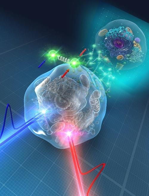
Caption: Molecular-vibrational imaging by stimulated Raman scattering microscopy utilizes two-color optical pulses to provide the vibrational spectroscopic signature of biomolecules
SRS is an interaction between two-color light and molecules. When the optical frequency difference of the two-color light (called as pump and Stokes) matches the molecular vibrational resonance frequency, the pump light is attenuated and the Stokes light is amplified as a result of SRS. To acquire SRS signal under a microscope, synchronized pump and Stokes pulse trains are generated, and Stokes pulse train is intensity-modulated in time. Then the pump and Stokes pulse trains are combined and focused on a sample. When SRS occurs, the intensity modulation of the Stokes pulse train is transferred to the pump pulse train. The transferred intensity modulation is detected by the lock-in detection technique to obtain the SRS signal. Images are acquired by scanning the stage or laser focal spot.
Different from CARS, SRS does not suffer from the non-resonant background (although there remain different mechanisms that contribute to a background of SRS), and therefore SRS image is easily interpretable because we can assume that SRS signal intensity is proportional to molecular concentration.
In the last decade, there has been tremendous progress on SRS microscopy. First, various methods of spectral imaging have been developed, where SRS images at various vibrational frequencies are acquired to discriminate different molecules. Second, various label-free imaging applications have been explored. Third, Raman probe imaging has been developed, where molecular tags are attached to biomolecules of interest, such as deuterium, alkyne and nitrile. So that we can monitor and track the metabolic activity, such as incorporation, biochemical reaction, and digestion of these biomolecules in cells. Super-multiplex imaging using Raman probes is also attracting attention. Lastly, the imaging modality is not limited to microscopy. The applications of SRS to endoscopy and flow cytometry have been successfully demonstrated, expanding the applications of vibrational imaging.
Although SRS microscopy may look mature, there remain various technical challenges such as the development of functional laser sources, functional Raman probes, and other methods for enhancing the sensitivity such as quantum optics. Such developments require interdisciplinary research including optics, laser engineering, quantum optics, chemistry, biology and medicine, and will lead to unexplored applications in biomedical imaging.