
Yuqing Xie, Yue Jing, Luyue Niu, Ci Wang, Lei Zhao, Jing Ren, Jianzhong Zhang. Perovskite-quantum-dots activated silica fiber X-ray dosimeter[J]. Chinese Optics Letters, 2022, 20(6): 063401
- Chinese Optics Letters
- Vol. 20, Issue 6, 063401 (2022)
Abstract
1. Introduction
X-ray detection technologies have played a crucial role in the fields of medical radiography, non-destructive inspection, high-energy physics research, etc.[
Recently, scintillators attached to the ends of polymethylmethacrylate (PMMA) plastic optical fibers have been implemented to obtain a real-time radiation dose monitoring (on-line dosimetry) at a precise point (punctual evaluation) very distant from the radiation source (remote monitoring)[
Scintillators based on rare earth (RE) ions emitted from 4f−4f forbidden transitions such as and are endowed with a long RL decay time (several milliseconds). On one hand, a long decay time is favorable for eliminating the Cherenkov interference, and, on the other hand, it sets an unfavorable limit to the sampling frequency[
Sign up for Chinese Optics Letters TOC. Get the latest issue of Chinese Optics Letters delivered right to you!Sign up now
The all-inorganic cesium lead halide (, ) perovskites have offered a promising option as a scintillator for X-ray imaging, owing to the following merits: high photoluminescence quantum yield (PLQY, ), fast radiative decay rate (a few tens of ns), good -ray stopping power, and tunable RL emission wavelengths (well matching a variety of photodetectors)[
However, PQDs still confront such an issue as a poor stability when exposed to light, heat, and moisture[
In this Letter, we constructed a new type of X-ray fiber dosimeter by coupling tellurite glass powders embedded with PQDs as the scintillator with a commercial multimode silica fiber (MMF) as the signal transmission waveguide [see the schematic diagram in Fig. 1(a)]. Experiments have verified that such a fiber dosimeter has a good linear responsiveness and hardness under high-dose X-ray irradiation, as well as a good stability when exposed to high temperature and water erosion. The spatial resolution of X-ray beam intensity is also studied using the proposed fiber sensor.
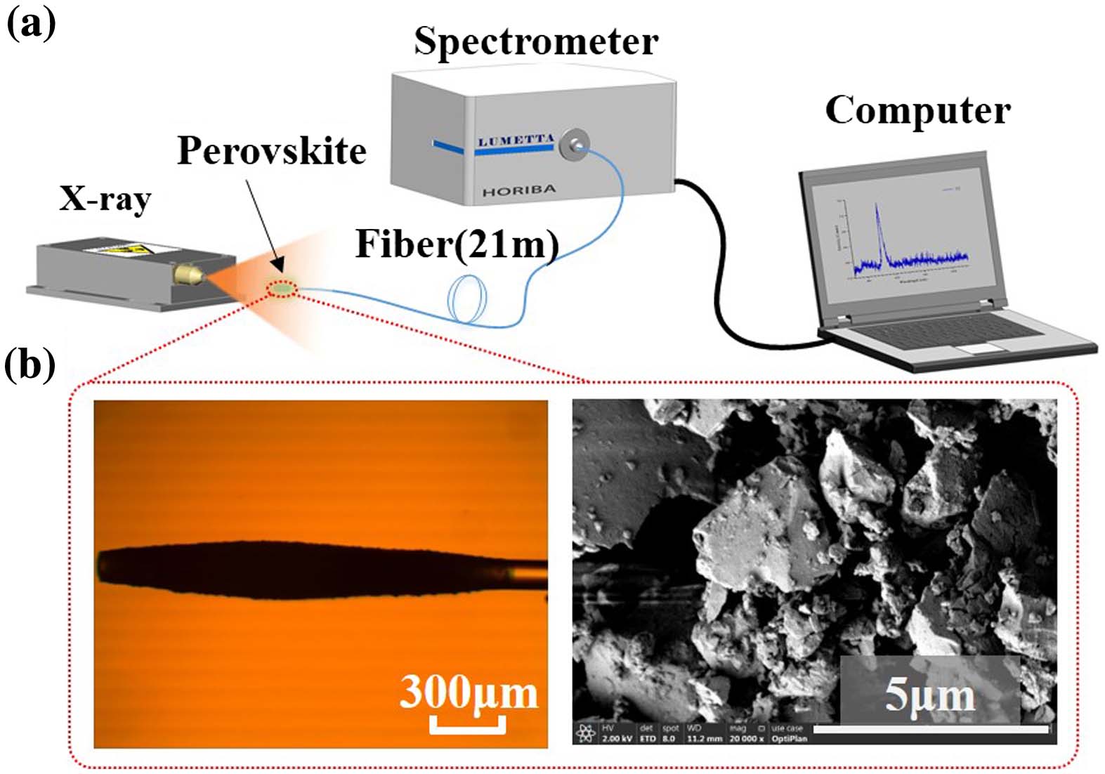
![]()
Figure 1.(a) Schematic diagram of the proposed X-ray detection set-up. (b) Microscopic image of the fiber head covered by PQDs embedded glass powders (left) and scanning electron microscope image of the glass powders (right).
2. Experiments and Methods
The PQDs embedded tellurite bulk glass was prepared by the conventional melt-quenching and subsequent thermal treatment method. The detailed preparation procedure can be found elsewhere[
The micromorphology of powdered glass samples was examined by scanning electron microscopy (SEM, Apreo S, Thermo Scientific). Photoluminescence (PL) emission, PL excitation (PLE), and decay spectra were measured by a PTI QuantaMaster 8000 spectrofluorometer (Horiba, Canada) equipped with a 75 W xenon lamp and a ns pulsed OpoLette355 optical parametric oscillator (OPO) laser (OPOTEK, Canada). X-ray excited RL spectra were recorded by an Omni-A 300i fluorescence spectrometer (Zolix, China) equipped with an X-ray tube (MOXTEK TUB-MAN-1006, the working voltage is up to 60 kV and 12 W, Cu target).
The manufacture of the X-ray fiber sensor is as follows: (i) prepare PQDs embedded glass powders by mechanically grinding the bulk glass in an agate mortar, and the powders thus obtained are of irregular shape and several tens of micrometers in size, as shown in Fig. 1(b); (ii) remove the polymer package from one end of a 21 m long MMF fiber, leaving a 0.2 cm long stripped fiber end; (iii) coat the fiber end with the glass powders using a transparent high-temperature glue (SINWE, UL-94V1, China) with a refractive index of 1.56, forming the X-ray sensitive film of in thickness [Fig. 1(b)]. The coated fiber is then used for X-ray radiation detection. During the measurement, the fiber sensor head was put 1 cm away from the radiation source. The other end of the fiber was connected to a CCD spectrometer (Horiba Lumett) for data reading.
The irradiation dose was expressed by Gy (air), and converted by multiplying the dose rate by the exposure time. The dose rate was measured without a filter by a multifunctional quality testing device of a Barakuda X-ray machine produced by RTI Company. The device consists of a main machine and a multifunctional detector (device number: BC1-08040132 + MPD-08070050). The dose rate was varied by the tube current, and the irradiation time (4 min) was precisely recorded during each run of measurements.
3. Results and Discussion
The PL emission, PLE, and decay spectra of PQDs embedded glass powders were characterized, as shown in Fig. 2. Upon the 365 nm UV excitation, a bright green emission is observed peaking at 525 nm with a narrow bandwidth of , which owes its origin to the bright triplet exciton radiative relaxation of PQDs [Fig. 2(a)][
![]()
Figure 2.(a) PL and PLE spectra of PQDs embedded glass powders. (b) PL decay curve and the fit to the data. (c) Variation of emission spectra and intensity under water boiling treatment repeated six times. Inset: glass powders in a boiling water bath. (d) Variation of emission spectra and intensity under heating (up to 300oC)–cooling (down to room temperature) treatment repeated six times.
The stability of PQDs embedded glass powders against humidity was checked in a boiling water bath [inset in Fig. 2(c)]. The powders that had been water boiled for 30 min were then measured to compare the changes in the emission spectra and intensity. The whole procedure was repeated six times. As shown in Fig. 2(c), the emission intensity only slightly changes under the repeated measurements, and the deviation in intensity is less than 3.6%, pointing to a good stability again with water erosion. The thermal stability of the powders was also studied under heating (up to 300°C) and cooling (down to room temperature) cycles. The whole process was repeated six times, and the results are shown in Fig. 2(d). Both the emission spectra and the intensity barely change under such treatments, and the deviation in intensity is less than 1%.
The RL spectrum of the PQDs activated silica fiber was measured under X-ray irradiation. Like the UV excitation, a strong and narrow green emission band is observed peaking at 525 nm [Fig. 3(a)][
![]()
Figure 3.(a) X-ray excited RL spectra of the PQDs activated silica fiber (blue curve) and the un-coated fiber (red curve). Inset: schematic diagram of the fiber sensor. (b) Relationship between the integrated RL intensity and the X-ray dose. Solid line is the linear fit to the data. (c) Variation of the RL intensity as a function of displacement across the X-ray beams. Red line is the Gaussian fit to the data. Inset: an X-ray sensitive glass used to record the X-ray beam spot. The background is the image of an enlarged spot after X-ray irradiation. (d) Changes in the RL spectra and integrated intensity with X-ray exposure time.
Due to the fairly small Stokes shift of , the self-absorption is inevitable, which sets a fundamental limit to light propagation and amplification. In this work, we only covered a very short length () of the silica fiber by the thin film of . The light emitted by is transmitting in the silica fiber to the spectrometer. The fiber per se is transparent and has limited absorption at the emission wavelength of . It is noted that there exist some novel perovskite scintillators based on self-trapped excitons with large Stokes shift. For example, Cheng et al. proposed a single crystal with a Stokes shift as large as 236 nm[
To investigate the spatial resolution of X-ray beam intensity, the fiber sensor was fixed on a 3D translation stage and placed close to the exit aperture of the X-ray tube. The emitted X-ray beam spot has a spatial spread of in diameter, as recorded by an X-ray sensitive glass [Ag-doped phosphate glass[
Finally, the stability of the PQDs activated fiber is examined upon continuous X-ray irradiation. The RL spectrum was recorded every 5 min for up to a total of 20 min. The RL spectra hardly change after the prolonged X-ray exposure with a cumulative dose of 200 Gy. The deviation of the integrated RL intensity before and after the exposure is only about 0.6% [Fig. 3(d)], pointing to an excellent radiation hardness of the proposed fiber sensor.
4. Conclusion
A simple X-ray fiber sensor is constructed by coupling PQDs embedded tellurite glass powders with a silica fiber. Capitalizing on the encapsulation of PQDs in the inorganic glass matrix, such a fiber sensor exhibits an excellent thermal stability upon the repeated heating–cooling cycles and a good resistance to water erosion. Benefitting from the strong RL emission from PQDs, the fiber sensor shows a good linear response to X-ray dose and can be reliably used upon continuous X-ray irradiation to a total dose of 200 Gy. The fiber sensor is also endowed with a good spatial resolution of the X-rays intensity distribution, and the resolving power is limited only by the fiber diameter.
References
[1] L. Lu, M. Sun, Q. Lu, T. Wu, B. Huang. High energy X-ray radiation sensitive scintillating materials for medical imaging, cancer diagnosis and therapy. Nano Energy, 79, 105437(2021).
[2] M. Sytnyk, S. Deumel, S. F. Tedde, G. J. Matt, W. Heiss. A perspective on the bright future of metal halide perovskites for X-ray detection. Appl. Phys. Lett., 115, 190501(2019).
[3] K. Li, Y. Gao, H. Zhang, G. Du, H. Huang, H. Xu, T. Xiao. Efficient three-dimensional characterization of C/C composite reinforced with densely distributed fibers via X-ray phase-contrast microtomography. Chin. Opt. Lett., 19, 073401(2021).
[4] D. Pennicard, B. Pirard, O. Tolbanov, K. Iniewski. Semiconductor materials for X-ray detectors. MRS Bull., 42, 445(2017).
[5] M. Nikl, A. Yoshikawa. Recent R&D trends in inorganic single-crystal scintillator materials for radiation detection. Adv. Opt. Mater., 3, 463(2015).
[6] M. Jia, J. Wen, X. Pan, L. Zhang, J. Yuan, Y. Huang, X. Zhang, L. He, F. Pang, T. Wang. Flexible scintillation silica fiber with engineered nanocrystals for remote real-time X-ray detection. ACS Appl. Mater. Interfaces, 14, 1362(2022).
[7] B. Sun, Y. Xie, Y. Zhao, X. Li, J. Chen, Y. Song, L. Zhao, Z. Li, H. Zhao, J. Ren, J. Zhang. A highly robust Ce3+-doped and Gd3+-mixed KLaF4 nano-glass composite scintillator. J. Mater. Chem. C, 9, 17504(2021).
[8] H. El Hamzaoui, G. Bouwmans, B. Capoen, A. Cassez, R. Habert, Y. Ouerdane, S. Girard, D. Di francesca, N. Kerboub, A. Morana, D. Söderström, A. Boukenter, M. Bouazaoui. Gd3+-doped sol-gel silica glass for remote ionizing radiation dosimetry. OSA Contin., 2, 715(2019).
[9] J. M. Fontbonne, G. Iltis, G. Ban, A. Battala, J. C. Vernhes, J. Tillier, N. Bellaize, C. Le Brun, B. Tamain, K. Mercier, J. C. Motin. Scintillating fiber dosimeter for radiation therapy accelerator. IEEE Trans. Nucl. Sci., 49, 2223(2002).
[10] L. Ding, Q. Wu, Q. Wang, Y. Li, R. M. Perks, L. Zhao. Advances on inorganic scintillator-based optic fiber dosimeters. EJNMMI Phys., 7, 60(2020).
[11] Z. Qin, Y. Hu, Y. Ma, W. Zhao, W. Sun, D. Zhang, Z. Chen, L. Elfed. Embedded structure fiber-optic radiation dosimeter for radiotherapy applications. Opt. Express, 24, 5172(2016).
[12] M. D. Belley, O. Craciunescu, Z. Chang, B. W. Langloss, I. N. Stanton, T. T. Yoshizumi, M. J. Therien, J. P. Chino. Real-time dose-rate monitoring with gynecologic brachytherapy: results of an initial clinical trial. Brachytherapy, 17, 1023(2018).
[13] S. Girard, D. Di Francesca, A. Morana, C. Hoehr, P. Paillet, C. Duzenli, N. Kerboub, I. Reghioua, G. Li Vecchi, A. Alessi, O. Duhamel, M. Trinczek, E. Marin, A. Boukenter, Y. Ouerdane, J. Mekki, R. Garcia Alia, Y. Kadi, M. Brugger. X-Rays, γ-rays, and proton beam monitoring with multimode nitrogen-doped optical fiber. IEEE Trans. Nucl. Sci., 66, 306(2019).
[14] M. Jia, J. Wen, X. Pan, Z. Xin, F. Pang, L. He, T. Wang. Tapered fiber radiation sensor based on Ce/Tb:YAG crystals for remote γ-ray dosimetry. Opt. Express, 29, 1210(2021).
[15] S. Kodama, S. Kurosawa, M. Ohno, Y. Morishita, H. Usami, M. Hayashi, M. Sasano, T. Azuma, H. Tanaka, V. Kochurikhin, A. Yamaji, M. Yoshino, S. Toyoda, H. Sato, Y. Ohashi, K. Kamada, Y. Yokota, A. Yoshikawa, T. Torii. Fiber-read radiation monitoring system using an optical fiber and red-emitting scintillator for ultra-high-dose conditions. Appl. Phys. Express, 13, 047002(2020).
[16] M. A. Suarez, T. Lim, L. Robillot, V. Maillot, T. Lihoreau, P. Bontemps, L. Pazart, T. Grosjean. Miniaturized fiber dosimeter of medical ionizing radiations on a narrow optical fiber. Opt. Express, 27, 35588(2019).
[17] F. Zhou, Z. Li, W. Lan, Q. Wang, L. Ding, Z. Jin. Halide perovskite, a potential scintillator for X-ray detection. Small Methods, 4, 2000506(2020).
[18] C. Wang, H. Lin, Z. Zhang, Z. Qiu, H. Yang, Y. Cheng, J. Xu, X. Xiang, L. Zhang, Y. Wang. X-ray excited CsPb(Cl,Br)3 perovskite quantum dots-glass composite with long-lifetime. J. Eur. Ceram. Soc., 40, 2234(2020).
[19] L. Jiang, X. Luo, Z. Luo, D. Zhou, B. Liu, J. Huang, J. Zhang, X. Zhang, P. Xu, G. Li. Interface and bulk controlled perovskite nanocrystal growth for high brightness light-emitting diodes [Invited]. Chin. Opt. Lett., 19, 030001(2021).
[20] Q. Chen, J. Wu, X. Ou, B. Huang, J. Almutlaq, A. A. Zhumekenov, X. Guan, S. Han, L. Liang, Z. Yi, J. Li, X. Xie, Y. Wang, Y. Li, D. Fan, D. B. L. Teh, A. H. All, O. F. Mohammed, O. M. Bakr, T. Wu, M. Bettinelli, H. Yang, W. Huang, X. Liu. All-inorganic perovskite nanocrystal scintillators. Nature, 561, 88(2018).
[21] R. T. Williams, W. W. Wolszczak, X. Yan, D. L. Carroll. Perovskite quantum-dot-in-host for detection of ionizing radiation. ACS Nano, 14, 5161(2020).
[22] D. Yu, P. Wang, F. Cao, Y. Gu, J. Liu, Z. Han, B. Huang, Y. Zou, X. Xu, H. Zeng. Two-dimensional halide perovskite as β-ray scintillator for nuclear radiation monitoring. Nat. Commun., 11, 3395(2020).
[23] H. Zhang, Z. Yang, M. Zhou, L. Zhao, T. Jiang, H. Yang, X. Yu, J. Qiu, Y. Yang, X. Xu. Reproducible X-ray imaging with a perovskite nanocrystal scintillator embedded in a transparent amorphous network structure. Adv. Mater., 33, 2102529(2021).
[24] L. Niu, S. Wang, Z. Sui, Y. Song, L. Zhao, L. Liu, J. Ren, J. Zhang. Highly stable CsPbBr3 perovskite quantum dot-doped tellurite glass nanocomposite scintillator. Opt. Lett., 46, 3448(2021).
[25] W. Ma, T. Jiang, Z. Yang, H. Zhang, Y. Su, Z. Chen, X. Chen, Y. Ma, W. Zhu, X. Yu, H. Zhu, J. Qiu, X. Liu, X. Xu, Y. Yang. Highly resolved and robust dynamic X-ray imaging using perovskite glass-ceramic scintillator with reduced light scattering. Adv. Sci., 8, 2003728(2021).
[26] S. Cheng, A. Beitlerova, R. Kucerkova, E. Mihokova, M. Nikl, Z. Zhou, G. Ren, Y. Wu. Non-hygroscopic, self-absorption free, and efficient 1D CsCu2I3 perovskite single crystal for radiation detection. ACS Appl. Mater. Interfaces, 13, 12198(2021).
[27] L. Lian, M. Zheng, W. Zhang, L. Yin, X. Du, P. Zhang, X. Zhang, J. Gao, D. Zhang, L. Gao, G. Niu, H. Song, R. Chen, X. Lan, J. Tang, J. Zhang. Efficient and reabsorption-free radioluminescence in Cs3Cu2I5 nanocrystals with self-trapped excitons. Adv. Sci., 7, 2000195(2020).
[28] M. Iwao, H. Takase, D. Shiratori, D. Nakauchi, T. Kato, N. Kawaguchi, T. Yanagida. Ag-doped phosphate glass with high weathering resistance for RPL dosimeter. Radiat. Meas., 140, 106492(2021).

Set citation alerts for the article
Please enter your email address
© Copyright 2018-2021 | Chinese Laser Press. All Rights Reserved 沪ICP备15018463号-20