The occurrence of numerous diseases, including cancer, cardiovascular diseases, and degenerative diseases, is closely related to the specific high expression of GSH. For instance, in A549 human lung adenocarcinoma cells, the GSH concentration was approximately an order of magnitude higher than that in normal cells. Therefore, the development of highly sensitive GSH detection and imaging approaches has crucial clinical value for the diagnosis of related diseases and a better understanding of the pathogenesis of the disease. The development of highly sensitive deep imaging approaches that can achieve GSH-specific responses in tumor tissues is still urgently needed. Photoacoustic (PA) imaging technology, as a novel biomedical imaging approach, which combines the high sensitivity of optical imaging with the deep penetration capability (up to 10 cm) of ultrasonic imaging, has been favored by the field of biomedical imaging in the last two decades. However, the characteristic molecules of many major diseases have weak optical absorption in the optical window of biological tissue (NIR-Ⅰ, 650-950 nm; NIR-Ⅱ, 950-1700 nm), resulting in the inability to generate a strong enough signal under excitation light irradiation, so that it is impossible to Realize photoacoustic imaging. Thus, developing nanoprobes with specific optical absorption properties as exogenous contrast agents for photoacoustic imaging can enhance the photoacoustic signal, thereby greatly improving the imaging contrast. Presently, various nanomaterials have been developed as exogenous contrast agents for photoacoustic imaging, including noble metal nanoprobes, carbon-based nano-2D materials, and high molecular polymers. Most of these materials are not biologically responsive and cannot specifically respond to specific substances in cells; they often lack the specific ability to recognize diseases. In this study, the authors developed a photosensitive AgBr@PLGA nanoprobe that can specifically respond to highly expressed GSH in the tumor microenvironment and proposed a tumor-specific near-infrared second region (NIR-Ⅱ) photoacoustic imaging approach.
The synthesized photosensitive AgBr@PLGA nanoprobes can be passively targeted to tumor tissue and can generate optical latent images triggered by external white light LEDs. GSH in the tumor microenvironment can reduce these optical latent images, resulting in a considerable number of silver nanoparticles that demonstrate strong light absorption and sharp improvements in photoacoustic signal in the NIR-Ⅱ region, thereby realizing specific photoacoustic imaging of tumor tissue.
The authors characterize the morphology and optical properties of the synthesized nanoprobes, and confirm in vitro their photosensitivity and GSH response characteristics in response to externally triggered white light LEDs. The experimental findings show that the prepared nanoparticles have good biocompatibility and ultra-high sensitivity to external trigger light, and the photoacoustic signal is continuously strengthened as exposure time increases. The model demonstrated that the synthesized AgBr@PLGA nanoprobes can attain high-contrast tumor-specific imaging in vivo, demonstrating the synthesized photosensitive great application potential of nanoprobes in tumor-specific photoacoustic detection and diagnosis.
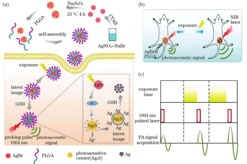
In this research, AgBr@PLGA nanocrystals were successfully used for ultrahigh-sensitivity and tumor-specific photoacoustic imaging through optical writing and redox chromogenic reactions. AgBr@PLGA NCs can show improved NIR-Ⅱ absorption because of the reduction of Ag nanoparticles when exposed to external trigger light after activation by GSH at the tumor site, as illustrated in Figure 2. The tumor-rich GSH content reduces the turn-on of NIR-II light absorption of AgBr@PLGA nanocrystals, enabling tumor-specific photoacoustic imaging with relatively high imaging depth, as demonstrated in Figure 5. Furthermore, this technique can accomplish contrast improvement in the tumor area by controlling the exposure time, and employing this approach can suppress unwanted background signals, such as blood signals in molecular imaging, as demonstrated in Figure 4. However, it should be noted that the response of the material at the imaging wavelength is not the same as the position of the absorption peak. We will develop photosensitive materials with strong absorption in the NIR-Ⅱ region in the following study to achieve deeper PA imaging. This study will attract more attention to the development of effective activatable PA probes for accurate biomedical imaging.