
Bin Ma, Jiaqi Han, Jing Li, Ke Wang, Shuang Guan, Xinshang Niu, Haoran Li, Jinlong Zhang, Hongfei Jiao, Xinbin Cheng, Zhanshan Wang. Damage characteristics of dual-band high reflectors affected by nodule defects in the femtosecond regime[J]. Chinese Optics Letters, 2021, 19(8): 081403
- Chinese Optics Letters
- Vol. 19, Issue 8, 081403 (2021)
Abstract
1. Introduction
In high-power laser systems, the damage resistance of optical components has received extensive attention. This characteristic directly affects the performance and life of the entire system. Several studies have shown that nodule defects remarkably reduce the laser-induced damage threshold (LIDT) of the optical coating[
A damage event is the result of a sequence of complex physical processes, including free carrier generation through multiphoton and avalanche ionization, and absorption and transmission of laser energy. For femtosecond laser pulses, stimulated nonlinear ionization is faster than the energy transfer between lattices and the formation of the temperature gradient field. The LIDT and related phenomenon are highly certain[
When designing the 800 nm (fs)/1064 nm (ns) dual-band high-reflection film, whether the main working band is designed in the inner layer or the outer layer needs to be considered. This condition determines whether the light energy is concentrated on the surface or inside the film. The damage mechanism becomes complicated due to the broadening of the incident angle and the electric field enhancement affected by different size nodules. This study mainly aims to explore the effects of artificial nodule defects with different sizes on the femtosecond laser damage behavior of dual-band high-reflection (HR) films. Combined with three-dimensional (3D) FDTD simulation and focused ion beam (FIB) profile images, a clear microscopic process and influencing factors of nodule damage are obtained, and the damage mechanism and damage behavior of the nodule in the femtosecond regime are clarified.
Sign up for Chinese Optics Letters TOC. Get the latest issue of Chinese Optics Letters delivered right to you!Sign up now
2. Experiments and Methods
A Ti:sapphire femtosecond laser system was used to provide 1 kHz, 800 nm laser pulses, and the pulse duration was approximately 50 fs () at the sample position. The Gaussian beam waist (1/e) at the location of the sample (vertical to the beam) was 94 µm (). The sample was positioned on a three-axis translation stage with positioning for the 45° AOI measurements by p-polarization. The LIDT test was implemented with R-on-1 process. At least 20 sites were tested for each sample, and the standard deviation of these LIDT statistics was taken as the error bar. Each single spot was irradiated by continuously rising energy starting from , with the maximum rising rate of shots. A high-magnification microscope was set to observe the surface of the sample and ensure that each nodule was moved to the center of the beam.
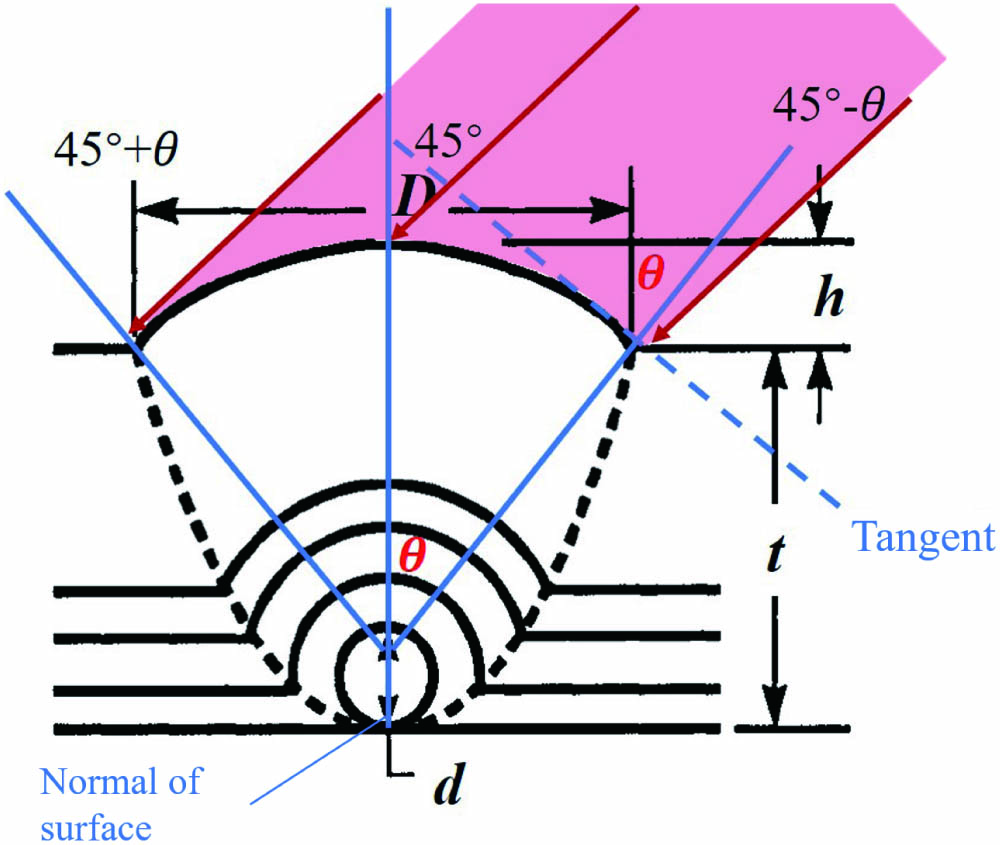
Monodisperse silica microspheres with diameters of 2.0, 1.5, 1.0, and 0.5 µm were deposited on the surface of the fused silica substrate. The density is about . An HR multilayer coating was deposited on the substrate {design: []}. H(h) and L (l) denote the high-refractive index (1.98) material () and low-refractive index (1.44) material (), respectively. The capital letters represent the working wavelength of 1064 nm, and h and l represent the working wavelength of 800 nm. Figure 1 shows schematic diagram of the nodule structure. The curve of the nodule surface is approximated as an arc. The real incident angle is to . The penetration position is on the edges of two sides of the nodule. Table 1 gives the calculated real incident angle range of different size nodules at 45° irradiation. Figure 2 shows the angle-dependent transmission curves of the HR film.
![]()
Figure 1.Schematic diagram of nodule structure.
| Size of Seeds | 2 µm | 1.5 µm | 1 µm | 0.5 µm |
|---|---|---|---|---|
| Range of AOI | 8.1°–81.9° | 12.8°–77.2° | 18.5°–71.5° | 26.1°–63.9° |
Table 1. Incidence Angle Range of Different Size Nodules
![]()
Figure 2.Angle-dependent transmission curve.
Figure 3(a) shows the 3D FDTD simulation results of the electric field inside the film under 45° p-polarized 800 nm laser irradiation for 2 µm seed nodules, with the boundary condition of a perfectly matched layer (PML). The simulation is based on the principle of , according to the actual profile measurement. The laser is incident from the left side, and the enhancement maximum occurs at two positions. The first position appears on the upper right side of the nodule structure, which is the peak value (the electric field is enhanced by 13 times), and the second position appears in the air on the right surface of the nodule (lower).
![]()
Figure 3.(a) FDTD-simulated |E2| distributions for the nodule. (b) |E2| along the coating without the nodule.
3. Results and Discussion
The blister is the common initial damage morphology of femtosecond laser-induced damage and is often generated at the position where the electric field is strongest in the film. The relationship among the laser damage induced by nodules, electric field enhancement, and blister generation is unclear due to the existence of nodules. Figure 4 shows the surface morphology and FIB profile of several damage states of the 2 µm seed nodule during the R-on-1 procedure. The position of damage can be obtained by comparing the FIB cross-sectional profile with the 3D FDTD simulation results. The damage sequence can be qualitatively described in the following way.
![]()
Figure 4.Damage state can be qualitatively described. (a) At low energy, rupture occurs on the side of the nodule that contacts the laser first. (b) Damage occurs at the place where the electric field is enhanced inside the nodule. (c) Modification of the film appears around the nodule. It is observed as a bright spot under a Nomarski microscope, which is the blister of film. (d) Large-scale catastrophic damage centered on the nodule.
First, as shown in Fig. 4(a), the film is peeled off on the left side surface of the nodule at low energy, although no internal damage is found. This condition is the blister damage of the nodule surface film, affected by the electric field of the designed film.
Subsequently, as shown in Fig. 4(b), damage occurs inside the nodule. The position is on the upper right side of the nodule, which is consistent with the FDTD simulation result. Although another maximum value of enhancement is found on the right side of the nodule surface, no damage occurs, perhaps because its position is displayed in the air. Electric field enhancement is the main way in which nodules affect the femtosecond laser damage. The initial damage process can be predicted through the simulation of the electric field to a certain extent. It is also related to the electric field intensity distribution in the film design, spectrum, range of incident angle, and structure stability. We infer that although the left side is one of the penetration areas, the light at this position actually reaches the electric field enhancement position on the right side less. Because it needs to pass a longer distance, and when light enters from the outermost air– interface, the incident angle will decrease, according to the law of refraction. Besides, the left surface contacts the laser first. These may cause the initial damage together. This order of occurrence of state 1 and state 2 requires further investigation.
Then, as shown in Fig. 4(c), the blister of the film appears near the nodule, which is the typical initial damage morphology of film. Affected by the nodule nearby, its morphology and LIDT are relatively unstable, which is different from ordinary HR films.
Finally, as shown in Fig. 4(d), the film exhibits layered destruction with the nodule as the center. The nodule is incompletely ejected. This situation is different from the nanosecond laser-induced damage incident at 45° and the femtosecond laser-induced damage incident at 0°.
The morphology of state 1 cannot be identified under an online microscope, so it is difficult to figure out what exactly the LIDT is. The profile of state 1 was obtained by FIB cutting after irradiation by laser energy, which is lower than the LIDT of state 2. Thereby, Table 2 and Fig. 5 show LIDT results of the second, third, and fourth states during the R-on-1 process. The LIDT of the nodule in state 1 or 2 is significantly lower than that of the film and is affected by the structural size of the nodule. The larger the size, the lower the LIDT. However, the LIDTs of the nodule and film in states 3 and 4 are extremely close. By contrast, the LIDT of state 4 for large-scale catastrophic damage shows an opposite trend. Specifically, the LIDT of the nodule is slightly higher than that of the HR film. This result is possibly because the incidence angle range is broadened because of the nodule. The larger the nodule size, the larger the range. Thus, more energy is deposited inside the film, and the energy on the surface is reduced. Therefore, more energy is needed to induce damage on the surface. Therefore, for state 4, the LIDT of larger nodules is shown to be higher.
![]()
Figure 5.LIDT results of each sample for R-on-1 process.
| LIDT ( | |||||
|---|---|---|---|---|---|
| 2 µm | 1.5 µm | 1.0 µm | 0.5 µm | HR Film | |
| State 2 | 0.309 | 0.377 | 0.481 | ||
| State 3 | 0.519 | 0.484 | 0.498 | 0.497 | 0.473 |
| State 4 | 0.532 | 0.515 | 0.512 | 0.509 | 0.487 |
Table 2. Measured LIDTs of Different Size Nodules and HR Film at States 2 to 4
As for the dual-band film, there should be a sequence of reflection designed to reflect two parts of wavelengths one after the other. Therefore, we reverse the order of the two parts of the HR film used in the above experiment. Specifically, the outer layer is the HR part of 1064 nm. At this time, the 800 nm laser directly penetrates this part and achieves HR inside the film. The mechanism of laser-induced damage is completely different. Under the same experimental conditions, the 2 µm seed nodule was tested with the R-on-1 procedure. The whole process roughly goes through five states, as shown in Fig. 6. Figure 7 shows the results of distribution and the FIB profiles of states 1 and 2. Table 3 gives the LIDTs.
| LIDT ( | |||||
|---|---|---|---|---|---|
| State | 1 | 2 | 3 | 4 | 5 |
| 2 µm seed nodule | 0.289 | 0.403 | 0.519 | 0.712 | 0.943 |
| HR film | 0.395 | 0.51 | 0.673 | 0.952 | |
Table 3. Measured LIDTs of the 2 µm Seed Nodules and the HR Film for the Second Type of Film
![]()
Figure 6.R-on-1 process mainly includes five states. 0. Initial nodule. 1. Rupture occurs on the nodule surface. This condition is observed as a change in scattered light under an online microscope. 2. A slight blister appears in the region of the light spot with the nodule as the center, and the height is approximately 30-50 nm. 3. The blister becomes serious, and its height reaches the micron level. 4. Limited growth on the surface near the nodule accompanied by the expansion of the blister area. 5. Growth to the inner layer.
![]()
Figure 7.(a) FDTD-simulated |E2| distribution for the nodule. (b) Electric field distribution along the coating. (c) FIB profile of state 1. (d) FIB profile of state 2.
Similarly, the nodule is still damaged before the surrounding film. However, the damage first appears inside the nodule, where the electric field is strongest. Most of the light is concentrated in the interior through the surface. That explains why the electric field enhancement level at the light converging area caused by the nodule is up to 60 times (13 times in the previous film). The film damage first occurs at the interface of the dual-band film structure, as shown in Fig. 7(d). It causes the entire film to undergo delamination and fracture, which is shown as a blister. In other words, the nodule damage caused by electric field enhancement has priority over the internal blister damage determined by the film design. With the increase of laser energy, the blister damage becomes severe, and a large area of delamination occurs, as shown in states 3–5 in Fig. 6.
In summary, our work verifies that a nodule defect is still an important factor affecting the femtosecond LIDT of optical films. The larger the nodule size, the lower the LIDT. Further studies have shown that the location of the femtosecond laser initial damage of nodules is incompletely determined by the position of electric field enhancement and is closely related to the process of blister damage. For high-reflection films with 45° incidence, FDTD simulation result shows that the strongest electric field is located inside the nodule. However, most of the energy is concentrated on the nodule surface. Thus, the damage occurs first at this location. Combined with the studies of dual-band film, it can be considered that the origination and growth of damage are a complex process affected by many factors. When the femtosecond reflective layer is inside the film, the area where the electric field is the strongest is destroyed first, followed by the blister at the reflective location and the large area of growth destruction. This condition is relatively different from the femtosecond laser-induced damage of the 0° reflector and the nanosecond laser-induced damage of the 45° reflector. These studies have provided new content and reference for the study of femtosecond laser-induced damage mechanism and law.
References
[1] K. H. Guenther. Nodular defects in dielectric multilayers and thick single layers. Appl. Opt., 20, 1034(1981).
[2] X. B. Cheng, Z. X. Shen, H. F. Jiao, J. L. Zhang, B. Ma, T. Ding, J. T. Lu, X. D. Wang, Z. S. Wang. Laser damage study of nodules in electron-beam-evaporated HfO2/SiO2 high reflectors. Appl. Opt., 50, C357(2011).
[3] X. B. Cheng, J. L. Zhang, T. Ding, Z. Y. Wei, H. Q. Li, Z. S. Wang. The effect of an electric field on the thermomechanical damage of nodular defects in dielectric multilayer coatings irradiated by nanosecond laser pulses. Light Sci. Appl., 2, e80(2013).
[4] V. E. Gruzdev, A. S. Gruzdeva. Resonance increase of high-power laser field with nodule defects in multilayer optical coatings: theory and simulation. Proc. SPIE, 3263, 169(1998).
[5] X. B. Cheng, A. Tuniyazi, Z. Y. Wei, J. L. Zhang, T. Ding, H. F. Jiao, B. Ma, H. Q. Li, T. B. Li, Z. S. Wang. Physical insight toward electric field enhancement at nodular defects in optical coatings. Opt. Express, 23, 8609(2015).
[6] X. B. Cheng, T. He, J. L. Zhang, H. F. Jiao, B. Ma, Z. S. Wang. Contribution of angle-dependent light penetration to electric-field enhancement at nodules in optical coatings. Opt. Lett., 42, 2086(2017).
[7] Y. G. Shan, H. B. He, C. Y. Wei, S. H. Li, M. Zhou, D. W. Li, Y. A. Zhao. Geometrical characteristics and damage morphology of nodules grown from artificial seeds in multilayer coating. Appl. Opt., 49, 4290(2010).
[8] C. Y. Wei, K. Yi, Z. X. Fan, J. D. Shao. Influence of composition and seed dimension on the structure and laser damage of nodular defects in HfO2/SiO2 high reflectors. Appl. Opt., 51, 6781(2012).
[9] H. P. Ma, X. B. Cheng, J. L. Zhang, H. F. Jiao, B. Ma, Y. J. Tang, Z. L. Wu, Z. S. Wang. Effect of boundary continuity on nanosecond laser damage of nodular defects in high-reflection coatings. Opt. Lett., 42, 478(2017).
[10] L. Y. Xie, T. He, J. L. Zhang, H. F. Jiao, B. Ma, Z. S. Wang, X. B. Cheng. Improve the LIDT of high-reflection coatings by planarizing nodular defects. High Power Laser Particle Beams, 30, 092001(2018).
[11] B. Mangote, L. Gallais, M. Zerrad, F. Lemarchand, L. H. Gao, M. Commandré, M. Lequime. A high accuracy femto-/picosecond laser damage test facility dedicated to the study of optical thin films. Rev. Sci. Instrum., 83, 013109(2012).
[12] N. S. Shcheblanov, T. E. Itina. Femtosecond laser interactions with dielectric materials: insights of a detailed modeling of electronic excitation and relaxation processes. Appl. Phys. A, 110, 579(2013).
[13] G. Y. Wang, L. Jiang, J. Y. Sun, J. Hu, Q. S. Wang, M. Li, Y. F. Lu. Ultrafast dynamics of three types of simultaneous shockwaves and filament attenuation in femtosecond laser multi-pulse ablation of PMMA. Chin. Opt. Lett., 17, 081405(2019).
[14] Y. X. Liu, T. J. Wang, N. Chen, H. Guo, H. Y. Sun, L. Zhang, Z. Qi, Y. X. Leng, Z. S. Wang, R. X. Li. Simultaneous generation of controllable double white light lasers by focusing an intense femtosecond laser pulse in air. Chin. Opt. Lett., 18, 121402(2020).
[15] H. Zhang, F. T. Zhang, X. Du, G. P. Dong, J. R. Qiu. Influence of laser-induced air breakdown on femtosecond laser ablation of aluminum. Opt. Express, 23, 1370(2015).
[16] N. Siaulys, L. Gallais, A. Melninkaitis. Direct holographic imaging of ultrafast laser damage process in thin films. Opt. Lett., 39, 2164(2014).
[17] L. Gallais, D. B. Douti, M. Commandre, G. Bataviciute, E. Pupka, M. Sciuka, L. Smalakys, V. Sirutkaitis, A. Melninkaitis. Wavelength dependence of femtosecond laser-induced damage threshold of optical materials. J. Appl. Phys., 117, 223103(2015).
[18] L. Gallais, B. Mangote, M. Zerrad, M. Commandré, V. Sirutkaitis. Laser-induced damage of hafnia coatings as a function of pulse duration in the femtosecond to nanosecond range. Appl. Opt., 50, C178(2011).
[19] S. Melnikas, T. Tolenis, L. Smalakys, G. Batavičiūtė, S. Kičas. Enhancement of laser-induced damage threshold in chirped mirrors by electric field reallocation. Opt. Express, 25, 26537(2017).
[20] S. L. Chen, P. P. Gao, Y. A. Zhao, Y. Z. Wang, Z. Fang, Y. X. Leng, J. D. Shao. Thermal-dynamical analysis of blister formation in chirped mirror irradiated by single femtosecond lasers. Appl. Opt., 53, 3347(2014).
[21] L. Gallais, X. B. Cheng, Z. S. Wang. Influence of nodular defects on the laser damage resistance of optical coatings in the femtosecond regime. Opt. Lett., 39, 1545(2014).
[22] X. Zou, F. Y. Kong, Y. X. Jin, P. Chen, J. M. Chen, J. Xu, Y. L. Wang, Y. B. Zhang, J. D. Shao. Influence of nodular defect size on metal dielectric mixed gratings for ultra-short ultra-high intensity laser system. Opt. Mater., 91, 177(2019).

Set citation alerts for the article
Please enter your email address
© Copyright 2018-2021 | Chinese Laser Press. All Rights Reserved 沪ICP备15018463号-20