Objective The technique called flow-mediated NADH fluorescence measurement is used to reflect tissue microcirculation function, which has important value for early screening of cardiovascular disease. However, due to the obvious difference in the extinction coefficients of oxyhemoglobin and deoxyhemoglobin in the visible and near-infrared bands, the tissue absorption coefficient will change constantly during the blood flow-mediated process. The excitation light and emission fluorescence of fluorescent molecules are affected by the tissue absorption coefficient. The current flow-mediated tissue fluorescence detection technology only measures the change in total tissue fluorescence intensity during brachial artery occlusion and release and does not account for the interference of tissue absorption coefficient changes caused by changes in oxygen saturation on NADH fluorescence measurement. Therefore, this problem may directly lead to blood flow-mediated tissue fluorescence technology failing to achieve an accurate measurement, thus limiting the clinical application of the technology.
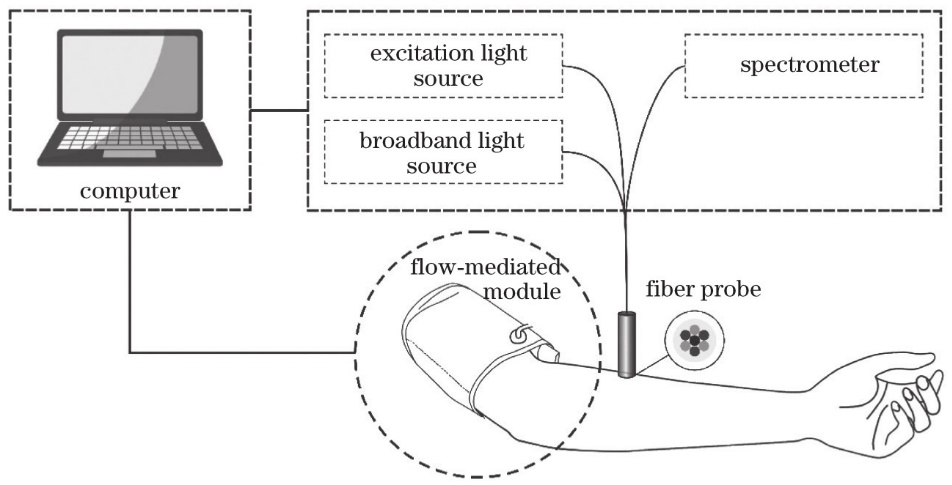
Methods First, we created the flow-mediated tissue fluorescence measurement system. The tissue absorption coefficient was calculated using a combination of tissue fluorescence and diffuse reflectance measurements. To improve the detection system’s accuracy, a steady-state tissue intrinsic fluorescence recovery method was used to correct the interference of absorption coefficient changes during the process of brachial artery occlusion and release on NADH fluorescence measurement. Second, we carried out the validation experiments of biological tissue solid phantom with different optical parameters and blood oxygen phantom simulating the physiological process of brachial artery occlusion and release. Furthermore, we validated the flow-mediated tissue fluorescence measurement system’s accuracy by comparing the corrected and uncorrected changes in blood flow-mediated tissue fluorescence in normal subjects.
Results and Discussions The results of tissue solid phantom experiments with different optical parameters showed when the values of α and β are 0.67 and 0.31, respectively, the small coefficient of variation of fluorescence intensity of the same concentration, the large linear correlation coefficient of gradient fluorescence intensity and concentration, and the closest curve shape could be satisfied at the same time (Fig.2). After obtaining the optimal combination of α and β , the fluorescence spectra of tissue phantom were recovered. The fluorescence intensity of NADH after recovery was linearly correlated with its concentration (R2=0.99), which indicated that the recovery effect was better (Fig.3). The results of blood oxygen phantom experiments showed that with the increase of sodium sulfite treatment time, oxygen saturation decreased significantly until stable. After being recharged, O2 returned to its baseline level and then decreased for the second time. The results demonstrated that the system could accurately extract the physiological parameters of a blood phantom. We found that changes in tissue oxygen saturation could interfere with NADH fluorescence detection, whereas the corrected intrinsic fluorescence spectrum of NADH was unaffected by changes in oxygen saturation (Fig.5). Finally, the blood flow-mediated tissue fluorescence system was used to measure the diffuse reflectance and fluorescence of four subjects during the brachial artery occlusion and release process. The low flow response (LFR) and high flow response (HFR) of four subjects were calculated separately. The results showed that the average values of LFR and HFR were 19.8% and 13.6%, respectively. In vivo experiments showed that after spectral recovery, the LFR and HFR decreased by 22.8% and 22.1%, respectively (Table 1). This change could be explained by the interference of oxygen saturation on the measured NADH fluorescence.
Conclusions The extraction of tissue optical parameters and the recovery of intrinsic fluorescence in steady-state tissue fluorescence technology were introduced into flow-mediated tissue fluorescence measurement in this study to achieve the dynamic measurement of tissue intrinsic fluorescence spectrum in the blood flow-mediated process. The tissue phantom experiment of the distribution of different absorption, scattering, and gradient fluorescence characteristics was validated using the flow-mediated tissue fluorescence and diffuse reflectance measurement system. The results showed that the coefficient of variation of fluorescence intensity of tissue phantom with the same concentration of fluorescence components was approximately 36% under different absorption and scattering characteristics, whereas the coefficient of variation of intrinsic fluorescence intensity was less than 10%. The results demonstrated that the recovery algorithm reduced the impact of absorption and scattering properties on the detection of the intrinsic fluorescence spectrum. Furthermore, there was a significant positive linear correlation between the intensity of the intrinsic tissue fluorescence spectrum and the concentration of fluorescent components, indicating that the intrinsic tissue fluorescence spectrum could be used to detect fluorescent components quantitatively. The results of blood oxygen phantom experiments showed that the change of blood oxygen saturation would interfere with the fluorescence measurement, and the corrected tissue intrinsic fluorescence spectrum was independent of the change of oxygen saturation, which further verified the reliability of the algorithm. Finally, it was found through in vivo experiments that the blood flow-mediated tissue fluorescence may better reflect changes in NADH fluorescence in tissues by introducing the tissue optical parameters extraction and tissue intrinsic fluorescence spectrum recovery algorithm, which was expected to effectively improve the accuracy of this technology’s tissue microcirculation function evaluation.