Hyperspectral microscopic imaging (HMI) technology combines optical microscopy and hyperspectral imaging to obtain both image and spectral information, thereby revealing spatial distribution and physical and chemical properties of a sample simultaneously. HMI, a novel nondestructive optical imaging technology, can be used to diagnose normal/cancerous tissues with high accuracy, sensitivity, and specificity. However, HMIs have a large amount of data and a complex data structure; thus, systematic and detailed data interpretation is required in cancer diagnosis. In this study, a push-broom HMI system is designed and developed, and the graphical user interface (GUI)-based software for system control, data acquisition, and data analysis is programmed to aid doctors in pathological diagnosis. The classification and staging of skin cancers (basal cell carcinoma, squamous cell carcinoma, and malignant melanoma) are studied on the basis of HMI technology and machine learning algorithms to confirm the performance of the system software. We hope that our HMI system, GUI-based software, and experimental results will be useful in cancer diagnosis and have application potential in biomedicine.
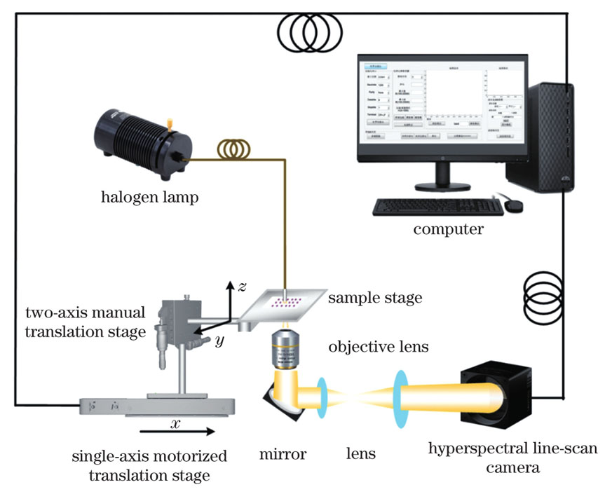
First, a push-broom HMI system consists of a halogen lamp, objective lens, sample stage, single-axis motorized translation stage, two-axis manual translation stage, hyperspectral line-scan camera, and other optical devices (Fig. 1). The halogen lamp illuminates the sample on the sample stage. The transmitted light is collected by the objective lens and directed to the hyperspectral camera after passing through the mirror and lens group in sequence to obtain one-dimensional (1D) spatial and spectral information. The motorized translation stage controls the sample stage to move in the x-direction with a step size of 1 μm for HMI data cube acquisition. The spectral resolution of the hyperspectral camera is calibrated and calculated based on the sensor configuration (Fig. 2). HMI system performance parameters, such as spatial resolution, field of view, and magnification, are obtained by imaging a resolution target. Second, the software with graphical user interfaces for system control, data acquisition, and data analysis is programmed using MATLAB. Several machine learning-based data processing methods are provided. Finally, the HMI data cubes of basal cell carcinoma, squamous cell carcinoma, and malignant melanoma tissues are obtained using the HMI system and data acquisition software; subsequently, the classification and staging of skin cancer are studied using data analysis software.
The push-broom HMI system has a spectral range of 465.5-905.1 nm, with a spectral resolution of ~3 nm, field of view of 400.18 μm×192.47 μm, system magnification of 28.15, and actual spatial resolution of 1.10-1.38 μm (Fig.3); it can collect a data cube of 2048 pixel×985 pixel×151. Additionally, GUI-based HMI data acquisition software and analysis software are designed and programmed using MATLAB. The data acquisition software includes the following three modules (Fig. 4): HMI system control and data acquisition module for controlling the hyperspectral camera and motorized translation stage, HMI data acquisition, light source background correction, and frequency domain filtering; HMI data display and processing module for displaying or cropping the HMI data cube and single-band image and calculating the correlation between each band; and save and exit module for saving the data processing results and exiting the acquisition software. The data analysis software consists of the following two modules (Fig. 5): a data extraction and viewing module that can realize HMI image display, spectrum viewing in the region of interest, converting 3D HMI data into 2D spectral data, and synthesizing RGB images with any three single-band images; and an HMI data processing module that can analyze image and spectral data and realize sample classification based on machine learning. HMI data from basal cell carcinoma, squamous cell carcinoma, and malignant melanoma are obtained to evaluate the performance of the system, and the machine learning is used to achieve the classification of three types of skin cancers and staging of squamous cell carcinoma. Spectral distribution, as well as 3D HMI, single-band, and RGB images, can be displayed (Fig. 6). The classification of three types of skin cancers based on image data is achieved using the data analysis software, and the highest classification accuracy of 85% and KAPPA value of 0.77 are obtained using color moment, gray-level co-occurrence matrix and local binary pattern as image features, partial least squares for dimensionality reduction, hold-out method for dividing the dataset, and a support vector machine models for classification. The optimal model for spectral data staging of squamous cell carcinoma corresponding to the standard normal variable transformation for spectral preprocessing, partial least squares for dimensionality reduction, hold-out method for dividing the dataset, and random forest for staging. The highest staging accuracy of 96.4% and a KAPPA value of 0.95 are obtained (Table 1).
In this study, a push-broom HMI system that can simultaneously obtain image and spectral information is developed to reveal spatial distribution and physicochemical properties of the samples. The HMI system can provide a data cube of 2048 pixel×985 pixel×151, a spectral resolution of ~3 nm, and actual spatial resolution of 1.10-1.38 μm. The HMI data acquisition software and analysis software are programmed using MATLAB. The graphical user interface of the software can standardize experiment procedures, allows intuitive data collection and processing, and provides analysis results, all of which can assist doctors in pathological diagnosis. Using this HMI system to image skin cancer tissues, high spectral and spatial resolution images are obtained, and the classification of different skin cancers and staging of squamous cell carcinomas can be achieved with high accuracy using machine learning algorithms. Our study shows that the combination of HMI technology and machine learning has significant application potential in the field of biomedicine.