Author Affiliations
1Sci-Tech Archaeology Center, Laboratory of Micro-Nano Optoelectronic Materials and Devices, Key Laboratory of Materials for High-Power Laser, Shanghai Institute of Optics and Fine Mechanics, Chinese Academy of Sciences, Shanghai 201800, China2Center of Materials Science and Optoelectronics Engineering, University of Chinese Academy of Sciences, Beijing 100049, China3Henan Provincial Institute of Cultural Relics and Archaeology, Zhengzhou 450000, Chinashow less
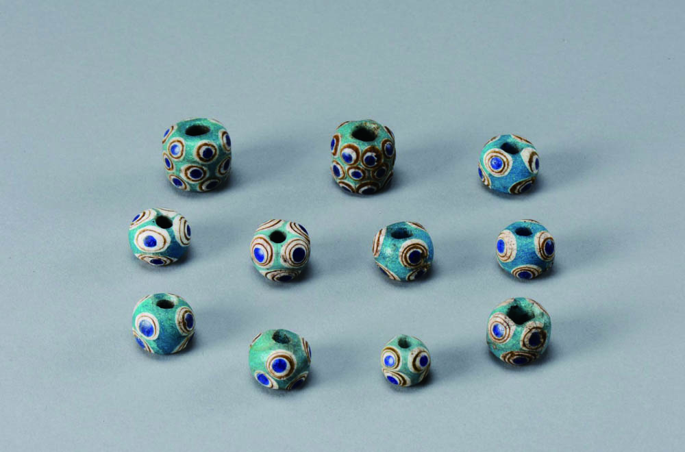
Fig. 1. Eye beads unearthed from Chu tomb in Xujialing, Xichuan, Henan Province.
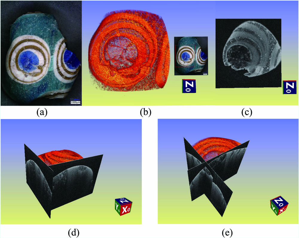
Fig. 2. Micrograph and 3D pseudo-color OCT images of the eye bead HXX-M10. (a) Micrograph of the broken bead; (b) top view and the position; (c) slice perpendicular to the axis, positioned at a distance of 184 pixels from the top; (d), (e) 3D sliced diagrammatic sketch at different angles along the x and y axes.
Fig. 3. 2D-OCT images of eye bead HXX-M10.
Fig. 4. Microscopic images of the fragments of eye bead HXX-M10.
Fig. 5. Element mapping for eye bead HXX-M10 shows that the dark blue area has a correlation with elements of Co and Fe, the white area with Sb and Ca, the brown area with Fe and Mn, and the blue matrix with Cu. The lighter shade corresponds to higher concentrations.
Fig. 6. , CuO, , CoO, and contents in different color regions of eye bead HXX-M10.
Fig. 7. Raman spectra of different color regions of eye bead HXX-M10. (a) Raman spectra of dark blue eye ball, turquoise blue matrix, and brown layer; (b) crystal in white circles; (c) envelope bands of turquoise blue matrix and crystal; (d) Oligoclase and crystals in a white layer. .
| Color | | MgO | | | Cl | | CaO | | | CoO | CuO | | | ZnO | | PbO | | SrO | BaO | |
|---|
| Mapping area | 9.78 | 0.63 | 4.14 | 70.56 | 0.45 | 0.84 | 9.29 | n.d. | 0.79 | 0.01 | 0.28 | 2.67 | 0.03 | n.d. | n.d. | n.d. | 0.46 | 0.03 | 0.03 | n.d. | | Dark blue | 1 | 7.79 | 0.58 | n.d. | 76.30 | 0.49 | 1.01 | 8.32 | 0.09 | 2.23 | 0.11 | 0.35 | 2.44 | n.d | 0.07 | 0.05 | 0.01 | 0.11 | 0.04 | n.d. | n.d. | | 2 | 15.81 | 0.35 | n.d. | 70.48 | n.d. | 0.77 | 7.44 | 0.07 | 1.94 | 0.10 | 0.28 | 2.29 | n.d | 0.06 | 0.09 | 0.01 | 0.27 | 0.03 | n.d. | n.d. | | 3 | 2.37 | 1.02 | 1.36 | 78.33 | n.d. | 1.24 | 9.73 | 0.12 | 2.51 | 0.13 | 0.42 | 1.94 | n.d. | 0.08 | 0.09 | 0.01 | 0.50 | 0.05 | 0.12 | n.d. | | 4 | 6.26 | 0.48 | n.d. | 77.01 | 0.68 | 0.07 | 9.06 | 0.11 | 2.48 | 0.13 | 0.40 | 2.47 | n.d. | 0.08 | 0.12 | 0.01 | 0.42 | 0.06 | 0.16 | n.d. | | 5 | 12.74 | 0.38 | n.d. | 73.27 | n.d. | 0.85 | 8.00 | 0.08 | 1.98 | 0.10 | 0.32 | 1.54 | n.d. | 0.06 | 0.10 | 0.01 | 0.37 | 0.04 | 0.17 | n.d. | | Average | 8.99 | 0.56 | 0.27 | 75.08 | 0.23 | 0.79 | 8.51 | 0.09 | 2.23 | 0.11 | 0.35 | 2.14 | | 0.07 | 0.09 | 0.01 | 0.33 | 0.04 | 0.09 | | | STD | 4.76 | 0.24 | 0.54 | 2.84 | 0.29 | 0.39 | 0.80 | 0.02 | 0.24 | 0.01 | 0.05 | 0.35 | | 0.01 | 0.02 | 0.00 | 0.13 | 0.01 | 0.08 | | | Turquoise blue | 1 | 8.75 | n.d. | 0.44 | 78.72 | 0.46 | 1.65 | 7.39 | 0.06 | 0.37 | n.d. | 1.04 | 0.35 | n.d. | n.d. | 0.19 | 0.06 | 0.41 | 0.04 | 0.08 | n.d. | | 2 | 20.07 | n.d. | 0.35 | 70.78 | 0.37 | 1.14 | 5.75 | 0.04 | 0.27 | n.d. | 0.72 | 0.10 | 0.01 | n.d. | 0.11 | 0.04 | 0.12 | 0.03 | 0.10 | n.d. | | 3 | 0.34 | n.d. | 0.39 | 86.44 | 0.47 | 1.46 | 7.84 | 0.08 | 0.41 | n.d. | 1.20 | 0.42 | n.d. | n.d. | 0.31 | 0.06 | 0.41 | 0.04 | 0.11 | n.d. | | Average | 9.72 | | 0.39 | 78.65 | 0.43 | 1.42 | 6.99 | 0.06 | 0.35 | | 0.99 | 0.29 | n.d. | n.d. | 0.20 | 0.05 | 0.31 | 0.04 | 0.10 | | | STD | 8.08 | | 0.04 | 6.39 | 0.04 | 0.21 | 0.90 | 0.02 | 0.06 | | 0.20 | 0.14 | 0.00 | n.d. | 0.08 | 0.01 | 0.14 | 0.00 | 0.01 | | | Brown | 1 | 22.11 | n.d. | 1.24 | 65.61 | 0.45 | 1.16 | 6.12 | 0.15 | 0.76 | n.d. | 0.01 | 1.82 | 0.06 | 0.01 | 0.05 | 0.01 | 0.41 | 0.03 | n.d. | n.d. | | 2 | 2.41 | n.d. | 0.92 | 82.28 | 0.68 | 0.98 | 8.07 | 0.12 | 0.94 | n.d. | 0.06 | 2.83 | 0.07 | 0.02 | 0.08 | 0.01 | 0.33 | 0.04 | 0.15 | n.d. | | 3 | 1.53 | n.d. | 0.99 | 82.56 | 0.70 | 1.01 | 7.81 | 0.13 | 0.93 | n.d. | 0.02 | 3.15 | 0.07 | 0.02 | 0.10 | 0.01 | 0.65 | 0.05 | 0.27 | n.d. | | 4 | 1.35 | n.d. | 0.43 | 82.89 | 0.73 | 0.92 | 8.26 | 0.14 | 0.97 | n.d. | 0.02 | 2.84 | 0.08 | 0.02 | 0.09 | 0.01 | 1.06 | 0.05 | 0.14 | 0.02 | | Average | 6.85 | | 0.90 | 78.34 | 0.64 | 1.02 | 7.57 | 0.14 | 0.90 | | 0.03 | 2.66 | 0.07 | 0.02 | 0.08 | 0.01 | 0.61 | 0.04 | 0.14 | 0.01 | | STD | 8.82 | | 0.29 | 7.35 | 0.11 | 0.09 | 0.85 | 0.01 | 0.08 | | 0.02 | 0.50 | 0.01 | 0.00 | 0.02 | 0.00 | 0.28 | 0.01 | 0.06 | 0.00 | | White | 1 | 7.24 | 0.92 | 0.62 | 79.67 | 0.74 | 0.01 | 7.24 | 0.10 | 0.68 | n.d. | 0.01 | 2.09 | 0.05 | n.d. | 0.05 | 0.01 | 0.55 | 0.04 | n.d. | n.d. | | 2 | 0.49 | n.d. | 0.66 | 80.46 | 0.55 | 0.79 | 9.38 | n.d. | 0.54 | n.d. | 0.02 | 6.32 | 0.03 | n.d. | 0.09 | 0.02 | 0.61 | 0.05 | n.d. | n.d. | | 3 | 13.07 | n.d. | 0.69 | 75.46 | n.d. | 0.02 | 6.94 | n.d. | 0.46 | n.d. | 0.01 | 2.61 | 0.02 | n.d. | 0.10 | 0.02 | 0.55 | 0.04 | n.d. | n.d. | | 4 | 13.23 | 0.22 | n.d. | 71.58 | n.d. | n.d | 10.49 | n.d. | 0.37 | n.d. | 0.01 | 3.33 | 0.02 | n.d. | 0.06 | 0.03 | 0.44 | 0.04 | 0.19 | n.d. | | 5 | 0.12 | n.d. | 0.92 | 82.18 | n.d. | 0.06 | 8.13 | n.d. | 0.57 | n.d. | 0.04 | 6.81 | 0.03 | n.d. | 0.11 | 0.02 | 0.63 | 0.05 | 0.34 | n.d. | | 6 | 19.54 | n.d. | 0.28 | 68.26 | n.d. | 0.46 | 5.33 | n.d. | 0.28 | n.d. | 0.01 | 5.22 | 0.01 | n.d. | 0.03 | 0.01 | 0.38 | 0.03 | 0.14 | n.d. | | 7 | 0.13 | n.d. | 0.38 | 78.26 | n.d. | 0.62 | 8.32 | n.d. | 0.59 | n.d. | 0.04 | 10.46 | 0.02 | n.d. | 0.22 | 0.05 | 0.64 | 0.05 | 0.24 | n.d. | | 8 | 20.97 | n.d. | 0.79 | 64.94 | 0.39 | 0.47 | 7.36 | n.d. | 0.35 | n.d. | 0.01 | 3.94 | 0.02 | n.d. | 0.06 | 0.02 | 0.56 | 0.03 | 0.10 | n.d. | | Average | 9.35 | 0.14 | 0.54 | 75.10 | 0.21 | 0.30 | 7.90 | 0.01 | 0.48 | | 0.02 | 5.10 | 0.03 | | 0.09 | 0.02 | 0.55 | 0.04 | 0.13 | | | STD | 8.07 | 0.30 | 0.28 | 5.83 | 0.28 | 0.29 | 1.47 | 0.03 | 0.13 | | 0.01 | 2.57 | 0.01 | | 0.06 | 0.01 | 0.09 | 0.01 | 0.12 | | | Lower detection limit (ppm) | 350 | 150 | 100 | 50 | 10 | 10 | 10 | 1 | 1 | 1 | 1 | 1 | 1 | 0.1 | 1 | 0.5 | 10 | 0.1 | 1 | 0.1 |
|
Table 1. Chemical Composition of the Eye Glass Bead HXX-M10 Unearthed from Chu Tomb in Xujialing by μ-XRFa (wt%)