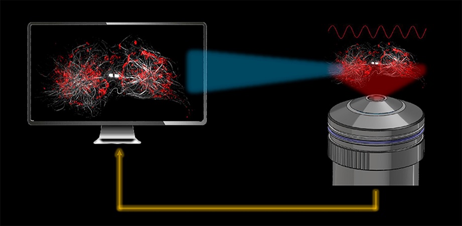
JSFR-SIM enables real-time observation of fine subcellular dynamics. Credit: Zhaojun Wang and Ming Lei, Xi'an Jiaotong University、
Superresolution structured illumination microscopy (SR-SIM) is an outstanding method for visualizing the subcellular dynamics in living cells. SR-SIM can achieve rapid, optically sectioned (OS), superresolution (SR) observation with hundreds to thousands of time points, which are the number of superresolution frames for continuous imaging.
However, the reconstruction algorithm for OS-SR-SIM imposes a significant computing burden, due to a complex workflow and a large number of calculations. This effectively nullifies real-time imaging, as 4 to 8 seconds are required to reconstruct a single SR image of 1024×1024 pixels. Microscope operators must first use the widefield mode to navigate the promising field-of-view for their investigations, then switch to the SR-SIM mode to acquire the raw images for SR-SIM, and then wait a long time to observe a single SR image, after dedicated postprocessing. The disjointed workflow of SIM setup hinders widespread application of SR-SIM among biologists.
Currently, parallel computing tools such as GPUs can be employed to accelerate the SR reconstruction, but the result is limited to a modest raw image size.
As reported in Advanced Photonics, researchers from Xi’an Jiaotong University (XJTU, China) and at the University of Nebraska (USA) recently developed a rapid reconstruction algorithm, “Joint Space and Frequency Reconstruction” (JSFR-SIM), to address both reconstruction speed and the limitation known as the missing cone problem. They combine a spatial domain processing technique with OS-SR-SIM implemented in the frequency domain, achieving improved image reconstruction speed plus suppression of the fuzzy background in images of thick cells.
The execution speed of the reconstruction in the GPU environment is 80 times faster than the widely used Wiener-SIM. Critically, the speed increase does not come at the expense of image quality. To demonstrate the method, they performed real-time live-cell observation of COS-7 cells, revealing insights into microtubule dynamics and rapid mitochondrial tubulation.

JSFR-SIM enables real-time visualization of microtubule and mitochondria dynamics. Credit: Wang et al., doi 10.1117/1.AP.4.2.026003.
The JSFR-SIM method makes locating and imaging a field-of-view at superresolution a facile and routine process, so that complex intracellular dynamics can be visualized in real time and resulting images quantitated. Compatible with various SIM modalities, it shares the same hardware setup as the conventional 2D-SIM, so it can be easily applied in many new SIM modalities—including AO-SIM, pSIM, and GI-SIM—to eliminate the performance bottleneck from computational burden, even when imaging thicker samples. This breakthrough promises to improve the work efficiency of biologists and facilitate SR-SIM as a routine tool in biomedical laboratories.
Read the open access article by Z. Wang et al., “High-speed image reconstruction for optically sectioned, super-resolution structured illumination microscopy,” Adv. Photon. 4(2) 026003 (2022) doi 10.1117/1.AP.4.2.026003.