
Lulin Fan, Tongjun Xu, Shun Li, Zhangli Xu, Jiancai Xu, Jianqiang Zhu, Baifei Shen, Liangliang Ji. Collimated gamma beams with high peak flux driven by laser-accelerated electrons[J]. High Power Laser Science and Engineering, 2023, 11(2): 02000e26
- High Power Laser Science and Engineering
- Vol. 11, Issue 2, 02000e26 (2023)
Abstract
1 Introduction
In recent years, laser-driven particle sources, such as electrons[1], ions[2] and neutrons[3], have been greatly developed due to their promising applications in high-energy density physics, nuclear physics, and cancer therapy treatment. Based on laser-accelerated electrons, gamma-ray radiations are also gaining increasing interest due to their ultra-high peak brilliance, short pulse duration and small beam size[4–7]. Such compact gamma-ray sources could pave the way for nuclear photonics, producing ultra-short neutron sources and medical isotopes[8], and radiography. In particular, the small beam size and large peak flux of the laser-generated gamma-ray sources can greatly improve the contrast and spatial resolution for nondestructive radiography compared to other approaches[9]. In strong-field quantum-electrodynamics, a promising approach to observe the Breit–Wheeler electron–positron pair production[10] in the linear or nonlinear regime is to collide laser-driven gamma photons with superintense lasers[11], X-ray radiations[12] or with each other. This requires the gamma beams to be collimated, guaranteeing high photon density in the collision region.
There are three main mechanisms to generate gamma-ray beams based on laser-driven energetic electrons in experiments: betatron radiation[13], inverse Compton scattering (ICS)[14,15] and bremsstrahlung radiation[4,5,16,17]. In general, betatron radiation produces gamma rays with photon energies from hundreds of keV to MeV when electrons oscillate in the laser-driven plasma bubble field. In ICS, the number of photons obtained by laser photons scattered by high-energy electrons is usually at the
A key to increase the photon yield in bremsstrahlung radiation is enhancing the number of relativistic electrons in laser–plasma accelerations. For instance[16], using picosecond laser pulses of relatively high pulse energy, plasma wakefield acceleration in the self-modulated regime produces
Sign up for High Power Laser Science and Engineering TOC. Get the latest issue of High Power Laser Science and Engineering delivered right to you!Sign up now
In this work, to obtain high-yield low-divergence gamma sources, we first produce a collimated high-charge electron beam through picosecond laser-driven self-modulated wakefield acceleration (SM-LWFA). Then it is sent to a high-Z target. The bremsstrahlung gamma-ray photons are measured with a high-resolution Compton-scattering spectrometer (CSS). The latter contains a gradual magnetic field to improve the energy resolution. The measured spectra are reproduced with GEANT4 simulations, suggesting a total photon number of
2 Experimental setup
The experiment was carried out on the SG-II UP picosecond experimental platform[19] at the Shanghai Institute of Optics and Fine Mechanics (SIOM). A schematic diagram of the experiment is shown in Figure 1. A linearly polarized laser pulse with a pulse duration of
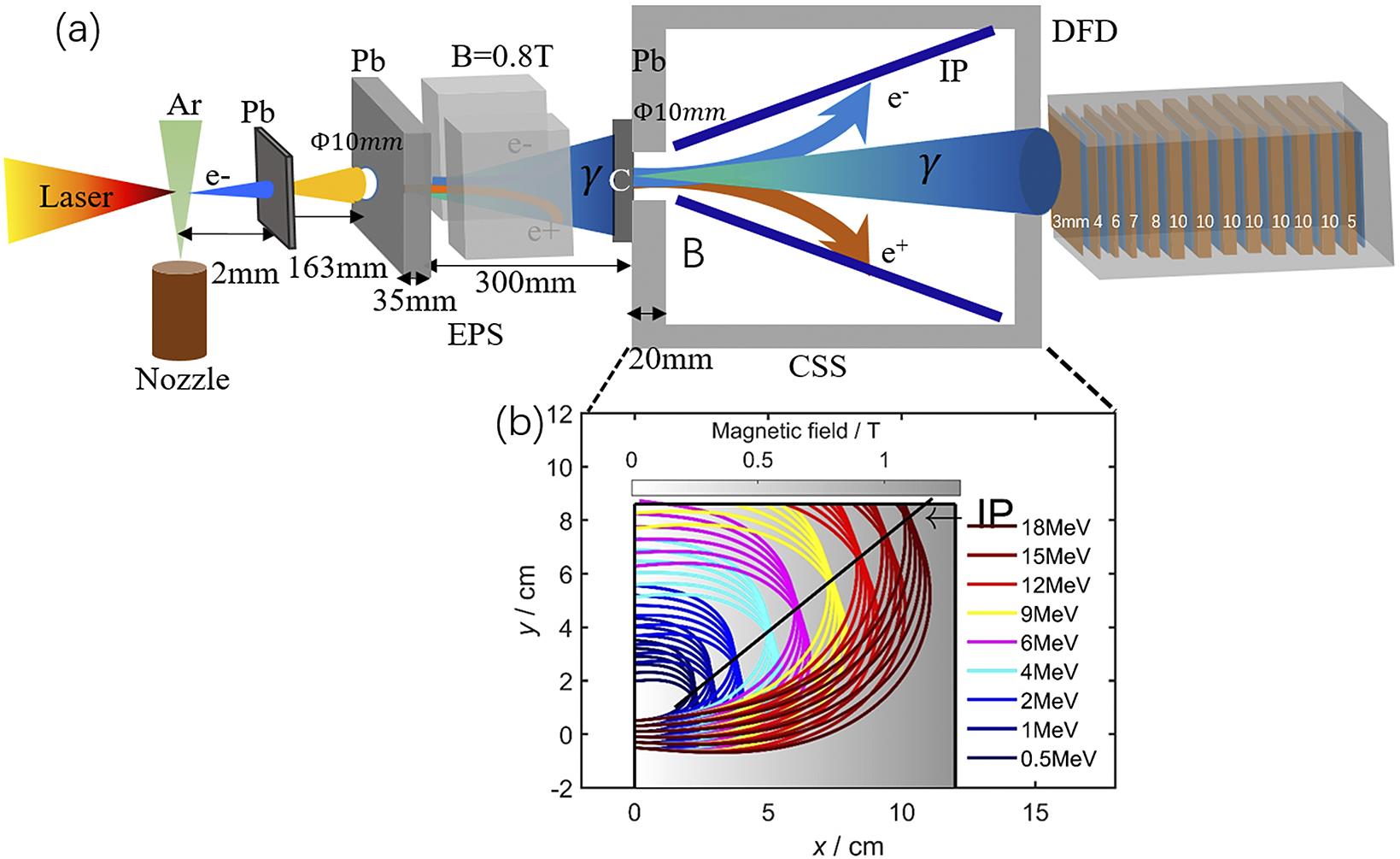
Figure 1.Schematic of the experimental setup. (a) A laser pulse propagates through an argon gas target, and energetic electrons are generated and collide with the 2 mm lead target located 2 mm behind the gas target to generate gamma-ray beams. An electron–positron spectrometer (EPS) with an aperture of 10 mm located  behind the lead target with an acceptance divergence angle of
  is added to deflect the positrons and electrons and measure their energy spectra. The gamma-ray beam spectra are measured with a typical differential filtering detector (DFD) and a Compton-scattering spectrometer (CSS) with a gradual magnet, which increases linearly along the laser direction and fills the whole spectrometer. The converter target in the CSS is carbon with thickness of 2 mm. The CSS and DFD are added
  behind the lead target, which has an acceptance divergence angle of
  . (b) Trajectories of the converted electron beams dispersed in the gradual magnetic field. These trajectories represent incident electron beams with energies of 0.5–18 MeV. The converted electrons enter the magnetic field with different transverse positions of
  and different angles of [–
 .
.
One key aspect of our experiments is the measurement of the gamma-ray spectrum. Gamma-ray beams driven by laser-accelerated electrons are of short pulse duration, comparable to that of the laser pulse (~1 ps here). Conventional scintillation and semiconductor detectors are not applicable to resolve the energy spectrum of the gamma-ray flash since all photons reach the detector in a short instance, resulting in the integrated photon energy being the sum of all received gamma-ray beam energy. Therefore, methods such as Compton scattering[22,23], photonuclear activation[24,25] and differential filtering[26,27] are employed to detect ultra-short gamma-ray flashes. The neutron separation thresholds relevant to the photonuclear activation cover a wide energy range[25], while the attenuation coefficients are not so sensitive to gamma energy above 2 MeV in differential filtering[28]. Thus, their spectrum resolutions are limited, especially in the high-energy region.
In this experiment, we chose a differential filtering detector (DFD) and a CSS together to detect these photons. A typical CSS usually uses a uniform magnetic field profile[22] or a stepped magnetic field profile[23] to deflect the photon-induced electron–positron pairs. The latter employs a curved surface plate to improve the energy resolution. Instead, we apply a gradual magnetic field for CSS, which increases linearly along the laser direction and fills the whole spectrometer. Thus, it is capable of gathering the converted electrons with the same energy but different emitting angles together and enhances the energy resolution of the gamma-ray beam, as shown in Figure 1(b). There is the electron–positron pair effect in the MeV gamma-ray range. The influence of the electron–positron pair effect can be largely eliminated through their mutual cancellation by the adoption of a symmetrical design for the spectrometer such that the positron and electron spectra are measured simultaneously. Then the energy spectrum of gamma beams can be obtained from the corrected converted electron energy spectrum. The DFD is placed behind the CSS, which consists of 13 pieces of lead filters with dimensions of
3 Experimental results and discussion
When a high-intensity
![]()
Figure 2.(a) Raw signal of the laser-accelerated electron beam recorded in the IP. (b) Extracted energy spectrum of the energetic electron beam. The black line represents the geometric mean value of the data of two shots. The shaded region represents uncertainty.
Removing the lead target and spectrometers, a spatial high-energy electron beam analyzer (SHEEBA)[32] composed of Al plates and IPs is located
![]()
Figure 3.(a)–(c) Spatial distribution of the electron beam recorded in the IP corresponding to different energies, namely,  MeV,
MeV,
These electrons with nC of charge and low divergence then collide with the 2-mm-thick lead target to generate gamma-ray photons through bremsstrahlung radiation. The EPS serves to remove the secondary electrons and positrons leaving the converting target here. The raw-data recorded by the DFD are shown in Figure 4(a) within the acceptance angle
![]()
Figure 4.(a) Raw-data of the gamma-photon signal recorded by the DFD. Raw-data of positrons (b) and electrons (c) recorded by the CSS. (d) Experimental spectra from the CSS (black solid), the DFD (red cross) and GEANT4 simulation with the experimental electrons as input (blue solid), within the divergence angle of  . These horizontal error bars represent 13 energy intervals and the vertical error bars represent uncertainty for the DFD. The black line represents the geometric mean value of the data and the shaded region represents uncertainty for the CSS.
To model the gamma-ray generation process, a series of test particle simulations are carried out with the Monte Carlo code GEANT4[36]. The simulation includes several physical processes, such as bremsstrahlung, scattering, ionization, pair production, photoelectric effect and Compton scattering. A total of
The simulated angular divergences of gamma-ray beams with different energies are summarized in Figure 5(a). It can be seen that the FWHM divergence of the gamma-ray beam of more than
![]()
Figure 5.(a) The divergence of gamma-ray beam by GEANT4 simulation with energy  . (b) Gamma-ray photon (
 ) yields and divergence (FWHM) versus different lead thicknesses. The simulation is performed with the experimental electrons as input.
The influence of thicknesses and the FWHM have also been studied by simulation, as shown in Figure 5(b). At the target thickness, the generated gamma-ray photon number increases due to continuous interaction between the electrons and the target. However, the energetic photons will be attenuated as the target thickness further increases. When these two processes reach a balance, the largest yield of gamma photons is obtained with the lead target thickness of
4 Conclusion
In conclusion, we use a picosecond laser to generate electron beams with large charge and low divergence, and subsequently to generate gamma-ray beams with high yield and low divergence through bremsstrahlung radiation. A typical DFD and a specially designed high detection resolution CSS with a gradual magnetic field are used at the same time to detect the generated gamma-ray beams precisely. The gamma-ray beams have a total photon number of
References
[1] A. J. Gonsalves, K. Nakamura, J. Daniels, C. Benedetti, C. Pieronek, T. C. H. de Raadt, S. Steinke, J. H. Bin, S. S. Bulanov, J. van Tilborg, C. G. R. Geddes, C. B. Schroeder, C. Toth, E. Esarey, K. Swanson, L. Fan-Chiang, G. Bagdasarov, N. Bobrova, V. Gasilov, G. Korn, P. Sasorov, W. P. Leemans. Phys. Rev. Lett., 122, 084801(2019).
[2] A. Macchi, M. Borghesi, M. Passoni. Rev. Mod. Phys., 85, 751(2013).
[3] M. M. Gunther, O. N. Rosmej, P. Tavana, M. Gyrdymov, A. Skobliakov, A. Kantsyrev, S. Zahter, N. G. Borisenko, A. Pukhov, N. E. Andreev. Nat. Commun., 13, 170(2022).
[4] Y. Glinec, J. Faure, L. L. Dain, S. Darbon, T. Hosokai, J. J. Santos, E. Lefebvre, J. P. Rousseau, F. Burgy, B. Mercier, V. Malka. Phys. Rev. Lett., 94, 025003(2005).
[5] A. Giulietti, N. Bourgeois, T. Ceccotti, X. Davoine, S. Dobosz, P. D’Oliveira, M. Galimberti, J. Galy, A. Gamucci, D. Giulietti, L. A. Gizzi, D. J. Hamilton, E. Lefebvre, L. Labate, J. R. Marques, P. Monot, H. Popescu, F. Reau, G. Sarri, P. Tomassini, P. Martin. Phys. Rev. Lett., 101, 105002(2008).
[6] X.-B. Wang, G.-Y. Hu, Z.-M. Zhang, Y.-Q. Gu, B. Zhao, Y. Zuo, J. Zheng. High Power Laser Sci. Eng., 8, e34(2020).
[7] G. Sarri, D. J. Corvan, W. Schumaker, J. M. Cole, A. Di Piazza, H. Ahmed, C. Harvey, C. H. Keitel, K. Krushelnick, S. P. Mangles, Z. Najmudin, D. Symes, A. G. Thomas, M. Yeung, Z. Zhao, M. Zepf. Phys. Rev. Lett., 113, 224801(2014).
[8] S. Janek, R. Svensson, C. Jonsson, A. Brahme. Phys. Med. Biol., 51, 5769(2006).
[9] J. C. Kieffer, A. Krol, Z. Jiang, C. C. Chamberlain, E. Scalzetti, Z. Ichalalene. Appl. Phys. B, 74, s75(2014).
[10] G. Breit, J. A. Wheeler. Phys. Rev., 46, 1087(1934).
[11] D. L. Burke, R. C. Field, G. HortonSmith, J. E. Spencer, D. Walz, S. C. Berridge, W. M. Bugg, K. Shmakov, A. W. Weidemann, C. Bula, K. T. McDonald, E. J. Prebys, C. Bamber, S. J. Boege, T. Koffas, T. Kotseroglou, A. C. Melissinos, D. D. Meyerhofer, D. A. Reis, W. Raggk. Phys. Rev. Lett., 79, 1626(1997).
[12] T. Nousch, D. Seipt, B. Kämpfer, A. I. Titov. Phys. Lett. B, 755, 162(2016).
[13] S. Cipiccia, M. R. Islam, B. Ersfeld, R. P. Shanks, E. Brunetti, G. Vieux, X. Yang, R. C. Issac, S. M. Wiggins, G. H. Welsh, M.-P. Anania, D. Maneuski, R. Montgomery, G. Smith, M. Hoek, D. J. Hamilton, N. R. C. Lemos, D. Symes, P. P. Rajeev, V. O. Shea, J. M. Dias, D. A. Jaroszynski. Nat. Phys., 7, 867(2011).
[14] S. Chen, N. D. Powers, I. Ghebregziabher, C. M. Maharjan, C. Liu, G. Golovin, S. Banerjee, J. Zhang, N. Cunningham, A. Moorti, S. Clarke, S. Pozzi, D. P. Umstadter. Phys. Rev. Lett., 110, 155003(2013).
[15] K. T. Phuoc, S. Corde, C. Thaury, V. Malka, A. Tafzi, J. P. Goddet, R. C. Shah, S. Sebban, A. Rousse. Nat. Photonics, 6, 308(2012).
[16] N. Lemos, F. Albert, J. L. Shaw, D. Papp, R. Polanek, P. King, A. L. Milder, K. A. Marsh, A. Pak, B. B. Pollock, B. M. Hegelich, J. D. Moody, J. Park, R. Tommasini, G. J. Williams, H. Chen, C. Joshi. Plasma Phys. Control. Fusion, 60, 054008(2018).
[17] S. Li, B. Shen, J. Xu, T. Xu, Y. Yu, J. Li, X. Lu, C. Wang, X. Wang, X. Liang, Y. Leng, R. Li, Z. Xu. Phys. Plasmas, 24, 093104(2017).
[18] S. Corde, K. T. Phuoc, G. Lambert, R. Fitour, V. Malka, A. Rousse, A. Beck, E. Lefebvre. Rev. Mod. Phys., 85, 1(2013).
[19] J. Zhu, J. Zhu, X. Li, B. Zhu, W. Ma, X. Lu, W. Fan, Z. Liu, S. Zhou, G. Xu, G. Zhang, X. Xie, L. Yang, J. Wang, X. Ouyang, L. Wang, D. Li, P. Yang, Q. Fan, M. Sun, C. Liu, D. Liu, Y. Zhang, H. Tao, M. Sun, P. Zhu, B. Wang, Z. Jiao, L. Ren, D. Liu, X. Jiao, H. Huang, Z. Lin. High Power Laser Sci. Eng., 6, e55(2018).
[20] T. Xu, B. Shen, J. Xu, S. Li, Y. Yu, J. Li, X. Lu, C. Wang, X. Wang, X. Liang, Y. Leng, R. Li, Z. Xu. Phys. Plasmas, 23, 033109(2016).
[21] G. J. Williams, B. R. Maddox, H. Chen, S. Kojima, M. Millecchia. Rev. Sci. Instrum., 85, 11E604(2014).
[22] D. J. Corvan, G. Sarri, M. Zepf. Rev. Sci. Instrum., 85, 065119(2014).
[23] Z.-C. Zhang, T. Yang, G.-Y. Hu, M.-T. Li, W. Luo, N. An, J. Zheng. Matter Radiat. Extremes, 6, 014401(2021).
[24] W. P. Leemans, D. Rodgers, P. E. Catravas, C. G. R. Geddes, G. Fubiani, E. Esarey, B. A. Shadwick, R. Donahue, A. Smith. Phys. Plasmas, 8, 2510(2001).
[25] M. M. Günther, K. Sonnabend, E. Brambrink, K. Vogt, V. Bagnoud, K. Harres, M. Roth. Phys. Plasmas, 18, 083102(2011).
[26] C. D. Chen, J. A. King, M. H. Key, K. U. Akli, F. N. Beg, H. Chen, R. R. Freeman, A. Link, A. J. Mackinnon, A. G. MacPhee, P. K. Patel, M. Porkolab, R. B. Stephens, L. D. Van Woerkom. Rev. Sci. Instrum., 79, 10E305(2008).
[27] R. Nolte, R. Behrens, M. Schnurer, A. Rousse, P. Ambrosi. Radiat. Protect. Dosim., 84, 367(1999).
[28] J. H. Hubbell, H. A. Gimm, I. Øverbø. J. Phys. Chem. Ref. Data, 9, 1023(1980).
[29] L. M. Chen, W. C. Yan, D. Z. Li, Z. D. Hu, L. Zhang, W. M. Wang, N. Hafz, J. Y. Mao, K. Huang, Y. Ma, J. R. Zhao, J. L. Ma, Y. T. Li, X. Lu, Z. M. Sheng, Z. Y. Wei. J. Gao, and J. Zhang, Sci. Rep., 3, 1912(2013).
[30] T. Bonnet, M. Comet, D. Denis-Petit, F. Gobet, F. Hannachi, M. Tarisien, M. Versteegen, M. M. Aleonard. Rev. Sci. Instrum., 84, 103510(2013).
[31] J. L. Shaw, M. A. Romo-Gonzalez, N. Lemos, P. M. King, G. Bruhaug, K. G. Miller, C. Dorrer, B. Kruschwitz, L. Waxer, G. J. Williams, M. V. Ambat, M. M. McKie, M. D. Sinclair, W. B. Mori, C. Joshi, H. Chen, J. P. Palastro, F. Albert, D. H. Froula. Sci. Rep., 11, 7498(2021).
[32] M. Galimberti, A. Giulietti, D. Giulietti, L. A. Gizzi. Rev. Sci. Instrum., 76, 053303(2005).
[33] C. Thaury, E. Guillaume, S. Corde, R. Lehe, M. Le Bouteiller, K. Ta Phuoc, X. Davoine, J. M. Rax, A. Rousse, V. Malka. Phys. Rev. Lett., 111, 135002(2013).
[34] J. L. Shaw, N. Lemos, L. D. Amorim, N. Vafaei-Najafabadi, K. A. Marsh, F. S. Tsung, W. B. Mori, C. Joshi. Phys. Rev. Lett., 118, 064801(2017).
[35] S. P. Mangles, A. G. Thomas, M. C. Kaluza, O. Lundh, F. Lindau, A. Persson, F. S. Tsung, Z. Najmudin, W. B. Mori, C. G. Wahlstrom, K. Krushelnick. Phys. Rev. Lett., 96, 215001(2006).
[36] S. Agostinelli, J. Allison, K. Amako, J. Apostolakis, H. Araujo, P. Arce, M. Asai, D. Axen, S. Banerjee, G. Barrand, F. Behner, L. Bellagamba, J. Boudreau, L. Broglia, A. Brunengo, H. Burkhardt, S. Chauvie, J. Chuma, R. Chytracek, G. Cooperman, G. Cosmo, P. Degtyarenko, A. Dell’Acqua, G. Depaola, D. Dietrich, R. Enami, A. Feliciello, C. Ferguson, H. Fesefeldt, G. Folger, F. Foppiano, A. Forti, S. Garelli, S. Giani, R. Giannitrapani, D. Gibin, J. J. Gómez Cadenas, I. González, G. Gracia Abril, G. Greeniaus, W. Greiner, V. Grichine, A. Grossheim, S. Guatelli, P. Gumplinger, R. Hamatsu, K. Hashimoto, H. Hasui, A. Heikkinen, A. Howard, V. Ivanchenko, A. Johnson, F. W. Jones, J. Kallenbach, N. Kanaya, M. Kawabata, Y. Kawabata, M. Kawaguti, S. Kelner, P. Kent, A. Kimura, T. Kodama, R. Kokoulin, M. Kossov, H. Kurashige, E. Lamanna, T. Lampén, V. Lara, V. Lefebure, F. Lei, M. Liendl, W. Lockman, F. Longo, S. Magni, M. Maire, E. Medernach, K. Minamimoto, P. M. de Freitas, Y. Morita, K. Murakami, M. Nagamatu, R. Nartallo, P. Nieminen, T. Nishimura, K. Ohtsubo, M. Okamura, S. O’Neale, Y. Oohata, K. Paech, J. Perl, A. Pfeiffer, M. G. Pia, F. Ranjard, A. Rybin, S. Sadilov, E. Di Salvo, G. Santin, T. Sasaki, N. Savvas, Y. Sawada, S. Scherer, S. Sei, V. Sirotenko, D. Smith, N. Starkov, H. Stoecker, J. Sulkimo, M. Takahata, S. Tanaka, E. Tcherniaev, E. S. Tehrani, M. Tropeano, P. Truscott, H. Uno, L. Urban, P. Urban, M. Verderi, A. Walkden, W. Wander, H. Weber, J. P. Wellisch, T. Wenaus, D. C. Williams, D. Wright, T. Yamada, H. Yoshida, D. Zschiesche. Nucl. Instrum. Methods Phys. Res. Sect. A, 506, 250(2003).
[37] A. Döpp, E. Guillaume, C. Thaury, A. Lifschitz, F. Sylla, J. P. Goddet, A. Tafzi, G. Iaquanello, T. Lefrou, P. Rousseau, E. Conejero, C. Ruiz, K. T. Phuoc, V. Malka. Nucl. Instrum. Methods Phys. Res. Sect. A, 830, 515(2016).

Set citation alerts for the article
Please enter your email address
© Copyright 2018-2021 | Chinese Laser Press. All Rights Reserved 沪ICP备15018463号-20