
Xin Huang, Jin He, Yiguang Jiang, Zhuocheng Chen, Xing Duan, Long Zhang. Structural and optical properties evolution in pressure-induced amorphization of metal-organic framework ZIF-8[J]. Chinese Optics Letters, 2022, 20(9): 091603
- Chinese Optics Letters
- Vol. 20, Issue 9, 091603 (2022)
Abstract
1. Introduction
Metal-organic frameworks (MOFs) have well-defined units containing both inorganic and organic constituent elements, rich topologies, easily tunable porous structures, and extremely high specific surface area[
Most MOFs materials are in a crystalline state. Over 60,000 crystalline MOFs materials have been identified from synthesis. In recent years, amorphous MOFs (aMOFs) have also gradually become a hot topic[
Zeolitic imidazolate framework-8 (ZIF-8) combines the advantages of a traditional zeolite structure and novel MOFs materials with high crystallinity, strong thermal stability, large specific surface area and pore volume, and high porosity[
Sign up for Chinese Optics Letters TOC. Get the latest issue of Chinese Optics Letters delivered right to you!Sign up now
In this Letter, we investigated the evolution in structural and optical properties of ZIF-8 crystals during pressure-induced amorphization. We carried out a battery of characterizations including powder X-ray diffraction (PXRD), scanning electron microscopy (SEM), adsorption measurement, second-harmonic signal measurement, and PL spectroscopy. The structural evolution of ZIF-8 crystals under pressure has been revealed. The optical properties of ZIF-8 during the crystal-amorphous transition, including second-harmonic generation (SHG) NLO properties, guest-host luminescence, for example, encapsulated with Eu cations, have been investigated in detail. The structure–optical properties relationship has been established to shed new light on the design of optical functional MOFs materials.
2. Experimental Method
2.1. Crystalline ZIF-8
A solid mixture of and 2-methylimidazole (H-MeIM) (0.250 g) was dissolved in 40 mL of dimethylformamide (DMF). Solution was poured into a 100 mL blue cap bottle and stirred for 30 min. The bottle was sealed, and solvothermal synthesis was carried out at 130°C for 48 h. After a complete reaction, the solution was allowed to cool to ambient temperature overnight. The resultant ZIF-8 crystals were filtered off the precursor solution and washed three times with 50 mL of DMF, followed by drying in an oven at 130°C for 12 h.
2.2. Crystalline Eu at ZIF-8
was dissolved in 10 mL of DMF, and the ZIF-8 crystals were poured into the solution and kept for 72 h. The crystals were filtered out of the solution, washed three times with 10 mL of DMF, and dried at 130°C for 12 h.
2.3. Amorphization of ZIF-8
Amorphization of the ZIF-8 crystals was done by the dry press molding method or cold isostatic pressing. When the applied pressure is lower than 50 MPa, we only used the hydraulic powder machine. The pressure was held at 20 MPa and 50 MPa for 5 min, respectively. When the pressure is higher than 50 MPa, the hydraulic press can no longer meet the demand, and we need to use a cold isostatic press. The pressure was held at 100 MPa and 200 MPa for 5 min, respectively.
X-ray diffraction (XRD) patterns were recorded using a Bruker D8 ADVANCE diffractometer with Cu Ka radiation (40 kV, 30 mA, 2 deg/min from 5 to 50 deg, ). The microstructure of the sample was observed using field emission SEM. A specific surface area was obtained using a Micromeritics ASAP 2020 instrument to measure the adsorption isotherm at 77 K based on the Brunauer–Emmett–Teller (BET) method. Prior to the adsorption and the mercury porosimetry analysis, all samples were evacuated overnight for 24 h at 120°C under vacuum. SHG measurement was performed with an MStarter 100 Ultrafast confocal microscope, in which the excitation wavelength was 1030 nm (Metatest Co., Ltd., China). The PL spectra were recorded on an Edinburgh FLS920 fluorescence spectrophotometer equipped with a Xe-900 lamp as the excitation source. The decay curves were collected using an FLS920 instrument with an nF900 flash lamp as the excitation source.
3. Result and Discussion
3.1. Amorphization of ZIF-8 crystals
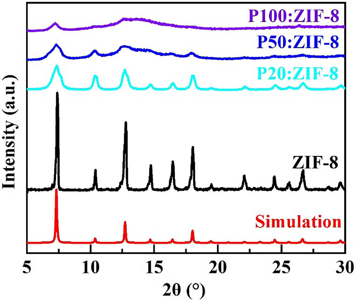
We promote amorphization of ZIF-8 crystals by continuously increasing pressure. The PXRD patterns of ZIF-8 crystals under different pressure clearly show the transition of ZIF-8 from the crystalline to amorphous state, as shown in Fig. 1. The characteristic diffraction peaks of the unpressurized ZIF-8 crystals were in good agreement with the XRD peaks of the simulated pattern formed from the crystals structure. The diffraction peaks are sharp, indicating that the prepared samples are ZIF-8 crystals with high purity and good crystallinity[
![]()
Figure 1.PXRD patterns of ZIF-8 under different pressure.
Under the pressure, the ZIF-8 crystals will gradually fragment into smaller powder particles. In order to observe the morphological changes of ZIF-8 crystals during amorphization, the crystals were characterized by SEM, as shown in Fig. 2. Figure 2(a) shows the morphology and distribution of the main elements of ZIF-8 crystals without stress. Grain growth is relatively regular, the grain size is about 10 µm, the aggregation of C, N, and Zn elements is relatively obvious, and the aggregation corresponds to the location of the ZIF-8. After being subjected to 50 MPa pressure, the structure of the ZIF-8 crystal was destroyed, and the whole ZIF-8 crystal was crushed into particles of different sizes, as shown in Fig. 2(b). Because the ZIF-8 crystal particles still retain part of their structure, the Bragg peaks in Fig. 1 are smoothed, but do not completely disappear.
![]()
Figure 2.SEM images and elemental distribution of ZIF-8 (a) before and (b) after pressurization. The atomic percentage of each element is shown at the bottom right of the images.
We analyzed the porosity and Brunauer–Emmett–Teller (BET) surface areas of the prepared samples by using adsorption at 77 K, as shown in Fig. 3. The adsorption curve and desorption curve of the samples almost overlapped without significant hysteresis, and the shape belonged to the typical I adsorption-desorption isotherm[
ZIF-8 consists of 2-methylimidazolate coordinated to a tetrahedral-linked metal center , which results in a zeolite-like topology. ZIF-8 demonstrates a strong intrinsic SHG signal due to its non-centrosymmetric cubic I-43m space group symmetry[
![]()
Figure 3.N2 adsorption isotherms for ZIF-8 before (black) and after (blue) pressurization. Solid circles indicate adsorption, while hollow circles indicate desorption.
![]()
Figure 4.SHG signal of ZIF-8 under different pressure excited by IR laser radiation (1030 nm central wavelength, 150 fs pulse duration, 50 mW, 80 MHz repetition rate).
3.2. Amorphization of Eu at ZIF-8 crystals
Based on the guest-host luminescence approach, we prepared Eu at ZIF-8 to further investigate the evolution of the structural and optical properties of ZIF-8 crystals during the pressure-induced amorphization. The ions were filled into the pores of ZIF-8 by prolonged soaking. Eu ions have turned out to be a promising medium for efficient infrared emission with a perspective of application in the respective laser and optical amplifier devices[
To confirm whether ions had entered the pores of ZIF-8 crystals, an elemental scan of the sample was performed. From Fig. 5(a), we can see that ions were successfully immersed into ZIF-8. The crystal morphology is relatively regular in shape and still around 10 µm in size. After being subjected to a pressure of 50 MPa, Eu at ZIF-8 was also crushed into small irregular particles. As the structure was damaged, some of the ions were released. Figure 6(a) shows that there is no obvious change in the XRD peaks of ZIF-8 crystals after loading with ions. ions are mainly loaded into the pores of ZIF-8 and do not destroy the lattice structure. Moreover, because of the support of in the pores, the degree of amorphization of Eu at ZIF-8 is lower than that of ZIF-8 under the same pressure.
![]()
Figure 5.SEM images and elemental distribution of Eu at ZIF-8 (a) before and (b) after pressurization. The atomic percentage of each element is shown at the bottom right of the images.
![]()
Figure 6.(a) PXRD patterns and (b) N2 adsorption isotherms of Eu at ZIF-8 before and after pressurization.
Compared with the ZIF-8 crystals, the BET surface area of Eu at ZIF-8 is reduced to . This is caused by the entry of ions into the pores to occupy a definite pore volume. The BET area of Eu at ZIF-8 was reduced to , and the pore volume also decreased from to after pressure amorphization, as shown in Fig. 6(b). The changing trend of the adsorption of Eu at ZIF-8 after amorphization is consistent with that of the ZIF-8 sample.
In the matrix, the crystal field or chemical change around the rare-earth ions is closely related to the microenvironment around the rare-earth ions. A slight change can cause the luminescence intensity or spectral splitting of the rare-earth ions. The local environment of the luminescence center affects the structure and distribution of the observed lines[
![]()
Figure 7.PL spectra for Eu at ZIF-8 before (black) and after (blue) pressurization.
4. Conclusions
We have induced amorphization of ZIF-8 by pressure to investigate the evolution of the structure and optical properties of ZIF-8 crystals during pressure-induced amorphization. At 20 MPa, the crystalline ZIF-8 already showed a tendency to amorphize. During the amorphization process, with increasing pressure, the lattice structure of ZIF-8 was gradually destroyed, the pores collapsed, and the BET surface area decreased significantly. The non-centrosymmetric crystal structure of ZIF-8 is altered during the crystal-amorphous transition, resulting in the decrease of SHG signal intensity. The amorphization effect on the guest-host luminescence behavior of ZIF-8 has been explored as well, via encapsulation with cations, the luminescence of which would reflect the spatial symmetry evolution. Accompanied with the destruction of the crystal lattice and the spatial symmetry of Eu at ZIF-8, the evolution of Eu cations luminescence, including the decrease of PL intensity and the increase of R ratio, has been observed. This work showed that the change of the lattice field environment not only affects the NLO properties of ZIF-8 itself, but also the luminescence of the guest species. The structural evolution in ZIF-8 amorphization and the structure–optical properties relationship provide a new avenue for the development of optical functional MOFs materials.
References
[1] Y. Q. Tian, C. X. Cai, X. M. Ren, C. Y. Duan, Y. Xu, S. Gao, X. Z. You. The silica-like extended polymorphism of cobalt(II) imidazolate three-dimensional frameworks: X-ray single-crystal structures and magnetic properties. Chemistry, 9, 5673(2003).
[2] P. I. Saragi, T. Spehr, A. Siebert, T. F. Lieker, J. Salbeck. Spiro compounds for organic optoelectronics. Chem. Rev., 107, 1011(2007).
[3] H. Kim, S. Yang, S. R. Rao, S. Narayanan, E. A. Kapustin, H. Furukawa, A. S. Umans, O. M. Yaghi, E. N. Wang. Water harvesting from air with metal-organic frameworks powered by natural sunlight. Science, 356, 430(2017).
[4] C. Li, K. Wang, J. Z. Li, Q. C. Zhang. Recent progress in stimulus-responsive two-dimensional metal–organic frameworks. ACS Mater. Lett., 2, 779(2020).
[5] J. A. Mason, J. Oktawiec, M. K. Taylor, M. R. Hudson, J. Rodriguez, J. E. Bachman, M. I. Gonzalez, A. Cervellino, A. Guagliardi, C. M. Brown, P. L. Llewellyn, N. Masciocchi, J. R. Long. Methane storage in flexible metal–organic frameworks with intrinsic thermal management. Nature, 527, 357(2015).
[6] M. S. Denny, J. C. Moreton, L. Benz, S. M. Cohen. Metal–organic frameworks for membrane-based separations. Nat. Rev. Mater., 1, 16078(2016).
[7] J. E. Mondloch, M. J. Katz, W. C. Isley, P. Ghosh, P. Liao, W. Bury, G. W. Wagner, M. G. Hall, J. B. DeCoste, G. W. Peterson, R. Q. Snurr, C. J. Cramer, J. T. Hupp, O. K. Farh. Destruction of chemical warfare agents using metal–organic frameworks. Nat. Mater., 14, 512(2015).
[8] H. J. Li, H. J. He, J. C. Yu, Y. J. Cui, Y. Yang, G. D. Qian. Dual-band simultaneous lasing in MOFs single crystals with Fabry–Perot microcavities. Sci China Chem., 62, 987(2019).
[9] Y. H. Wei, H. Y. Dong, C. Wei, W. Zhang, Y. L. Yan, Y. S. Zhao. Wavelength-tunable microlasers based on the encapsulation of organic dye in metal-organic frameworks. Adv. Mater., 28, 7424(2016).
[10] J. C. Yu, Y. J. Cui, C. D. Wu, Y. Yang, Z. Y. Wang, M. O. Keeffe, B. L. Chen, G. D. Qian. Second-order nonlinear optical activity induced by ordered dipolar chromophores confined in the pores of an anionic metal-organic framework. Angew. Chem. Int. Ed., 51, 10542(2012).
[11] S. Chen, S. Y. Yang, Y. Huang, W. Y. Jiao, G. H. Fan, Y. C. Gao. Wavelength-dependent nonlinear absorption of gold nanocages. Chin. Opt. Lett., 18, 011901(2020).
[12] M. M. Wang, M. K. Zhang, W. W. Song, L. Zhou, X. Y. Wang, Y. F. Tang. Heteroatom-doped amorphous cobalt–molybdenum oxides as a promising catalyst for robust hydrogen evolution. Inorg. Chem., 61, 5033(2022).
[13] A. C. Ghosh, A. Legrand, R. Rajapaksha, G. A. Craig, C. Sassoye, G. Balázs, D. Farrusseng, S. Furukawa, J. Canivet, F. M. Wisser. Rhodium-based metal–organic polyhedra assemblies for selective CO2 photoreduction. J. Am. Chem. Soc., 144, 3626(2022).
[14] H. Z. Wang, X. K. Pei, M. J. Kalmutzki, J. J. Yang, O. M. Yaghi. Large cages of zeolitic imidazolate frameworks. Acc. Chem. Res., 55, 707(2022).
[15] A. Qiao, T. D. Bennett, H. Z. Tao, A. Krajnc, G. Mali, C. M. Doherty, A. W. Thornton, J. C. Mauro, G. N. Greaves, Y. Z. Yue. A metal-organic framework with ultrahigh glass-forming ability. Sci. Adv., 4, 6827(2018).
[16] K. S. Park, Z. Ni, A. P. Côté, J. Y. Choi, R. D. Huang, F. J. Uribe-Romo, H. K. Chae, M. O. Keeffe, O. M. Yaghi. Exceptional chemical and thermal stability of zeolitic imidazolate frameworks. Proc. Natl. Acad. Sci. U.S.A., 103, 10186(2006).
[17] J. J. Ren, T. R. Li, X. P. Zhou, X. Dong, A. V. Shorokhov, M. B. Semenov, V. D. Krevchik, Y. H. Wang. Encapsulating all-inorganic perovskite quantum dots into mesoporous metal organic frameworks with significantly enhanced stability for optoelectronic applications. Chem. Eng. J., 358, 30(2019).
[18] G. N. Greaves, S. Sen. Inorganic glasses, glass-forming liquids and amorphizing solids. Adv. Phys., 56, 1(2007).
[19] T. D. Bennett, A. L. Goodwin, M. T. Dove, D. A. Keen, M. G. Tucker, E. R. Barney, A. K. Soper, E. G. Bithell, J. C. Tan, A. K. Cheetham. Structure and properties of an amorphous metal-organic framework. Phys. Rev. Lett., 104, 115503(2010).
[20] R. N. Widmer, G. I. Lampronti, S. Anzellini, R. Gaillac, S. Farsang, C. Zhou, A. M. Belenguer, C. W. Wilson, H. Palmer, A. K. Kleppe, M. T. Wharmby, X. Yu, S. M. Cohen, S. G. Telfer, S. A. T. Redfern, F.-X. Coudert, S. G. MacLeod, T. D. Bennett. Pressure promoted low-temperature melting of metal–organic frameworks. Nat. Mater., 18, 370(2019).
[21] A. S. Poryvaev, D. M. Polyukhov, M. V. Fedin. Mitigation of pressure-induced amorphization in metal-organic framework ZIF-8 upon EPR control. ACS Appl. Mater. Interfaces, 12, 16655(2020).
[22] M. Guo, H. B. He, K. Yi, S. Y. Shao, G. H. Hu, J. D. Shao. Optical characteristics of ultrathin amorphous Ge films. Chin. Opt. Lett., 18, 103101(2020).
[23] T. D. Bennett, A. K. Cheetham. Amorphous metal−organic frameworks. Acc. Chem. Res., 47, 1555(2014).
[24] D. F. Jimenez, R. Galvelis, A. Torrisi, A. D. Gellan, M. T. Wharmby, P. A. Wright, C. M. Draznieks, T. Düren. Flexibility and swing effect on the adsorption of energy-related gases on ZIF-8: combined experimental and simulation study. Dalton Trans., 41, 10752(2012).
[25] T. Tian, J. V. Garcia, T. D. Bennett, D. F. Jimenez. Mechanically and chemically robust ZIF-8 monoliths with high volumetric adsorption capacity. J. Mater. Chem. A, 3, 2999(2015).
[26] T. D. Bennett, S. Cao, J. C. Tan, D. A. Keen, E. G. Bithell, P. J. Beldon, T. Friscic, A. K. Cheetham. Facile mechanosynthesis of amorphous zeolitic imidazolate frameworks. J. Am. Chem. Soc., 133, 14546(2011).
[27] S. V. Cleuvenbergen, I. Stassen, E. Gobechiya, Y. X. Zhang, K. Markey, D. E. De Vos, C. Kirschhock, B. Champagne, T. Verbiest, M. A. Van Der Veen. ZIF-8 as nonlinear optical material: influence of structure and synthesis. Chem. Mater., 28, 3203(2016).
[28] Y. A. Mezenov, N. K. Kulachenkov, A. N. Yankin, S. S. Rzhevskiy, P. V. Alekseevskiy, V. D. Gilemkhanova, S. V. Bachinin, V. Dyachuk, V. A. Milichko. Polymer matrix incorporated with ZIF-8 for application in nonlinear optics. Nanomaterials, 10, 1036(2020).
[29] K. Driesen, V. K. Tikhomirov, C. Gorller-walrand. Eu3+ as probe for rare earth dopant site structure in nano-glass ceramics. J. Appl. Phys., 102, 024312(2007).
[30] X. M. Li, S. S. Zhou, R. F. Wei, X. Y. Liu, B. Q. Cao, H. Guo. Blue–green color-tunable emissions in novel transparent Sr2LuF7:Eu/Tb glass-ceramics for WLEDs. Chin. Opt. Lett., 18, 051601(2020).
[31] G. H. Jia, P. A. Tanner, C. K. Duan, J. Dexpert-Ghys. Eu3+ spectroscopy: a structural probe for yttrium orthoborate phosphors. J. Phys. Chem. C, 114, 2769(2010).
[32] D. V. Deyneko, I. V. Nikiforov, D. A. Spassky, Y. Y. Dikhtyar, S. M. Aksenov, S. Y. Stefanovich, B. I. Lazoryak. Luminescence of Eu3+ as a probe for the determination of the local site symmetry in β-Ca3(PO4)2-related structures. Cryst. Eng. Comm., 21, 5235(2019).

Set citation alerts for the article
Please enter your email address
© Copyright 2018-2021 | Chinese Laser Press. All Rights Reserved 沪ICP备15018463号-20